Article Text

Abstract
Background Vimentin is an intermediate-sized filament which is highly expressed in mesenchymal cells and is associated with epithelial–mesenchymal transition (EMT). EMT markers ZEB2 and Slug lead to Vimentin overexpression and E-cadherin loss, resulting in invasion and metastasis. However, the status of Vimentin remains unexplored in eyelid sebaceous gland carcinoma (SGC). The study aims to determine status of Vimentin in SGC and its association with EMT markers E-cadherin, ZEB2 and Slug.
Methods Vimentin protein expression was undertaken in 66 cases with SGC by immunohistochemistry (IHC). Messenger RNA (mRNA) expression was determined in 42 fresh tissues by quantitative real-time PCR. Association of Vimentin with E-cadherin, ZEB2 and Slug was also analysed. Patients were followed up for 17–69 months (mean 34.02 ± 14.73 months).
Results IHC revealed Vimentin overexpression in 37/66 (56%) cases. This overexpression showed significant association with lymph node metastasis (p=0.004) and pagetoid spread (p=0.05). Patients with high Vimentin expression also had poor disease-free survival (p=0.033). Univariate Cox regression model indicated that high Vimentin expression (p=0.043) and advanced tumour stage (p=0.002) were independent adverse prognostic factors. High Vimentin mRNA expression was seen in 16/42 (38%) cases and correlated significantly with lymph node metastasis (p=0.027), advanced tumour stage (p=0.002) and large tumour size (p=0.023). Vimentin expression overall showed a significant inverse association with E-cadherin and direct association with ZEB2 expression.
Conclusions Vimentin overexpression in SGC is associated with EMT and leads to poor clinical outcome. It also emerged as a novel predictor for lymph node metastasis and poor survival.
- Vimentin
- epithelial–mesenchymal transition
- eyelid sebaceous gland carcinoma
- quantitative real time PCR
- immunohistochemistry
Statistics from Altmetric.com
- Vimentin
- epithelial–mesenchymal transition
- eyelid sebaceous gland carcinoma
- quantitative real time PCR
- immunohistochemistry
Introduction
Sebaceous gland carcinoma (SGC) of the eyelid is a highly malignant and potentially lethal cutaneous neoplasm which arises in the glands of Zeis associated with hair follicles and from meibomian glands. It accounts for 27%–44% of all eyelid malignancies in Asian population.1 2 However, it is less common in Caucasian regions accounting for only 1%–5.5% of all eyelid tumours.3 4 It is known as the ‘great masquerader’, as it is often misdiagnosed, leading to a delay in diagnosis which contributes to an increase in associated morbidity and mortality.
Epithelial–mesenchymal transition (EMT) is a critical phenomenon in invasion, progression and metastasis of epithelial malignancies, which is characterised by loss of epithelial morphology and acquisition of a highly invasive mesenchymal phenotype.5 Decrease in the expression of cell adhesion molecule E-cadherin and increased expression of mesenchymal markers such as Vimentin along with aberrant expression of transcription factors (like ZEB2 and Slug) are considered as hallmarks of EMT.5 Vimentin, a 57 kDa protein, is an intermediate filament type III protein, that is expressed in normal mesenchymal cells to maintain cellular integrity and provide resistance against stress. It is expressed in a variety of cells including fibroblasts, endothelial cells, macrophages, neutrophils, leukocytes, etc. Overexpression of Vimentin in cancer correlates with tumour progression, metastasis and poor clinical outcome. It is overexpressed in various epithelial cancers including breast cancer, central nervous system (CNS) tumours, prostate cancer, malignant melanoma, lung cancer and gastrointestinal tumours.6 Transcription factors belonging to Snail family (Slug/Snail2) and ZEB family (ZEB2/SIP1) are considered to be master regulators triggering EMT leading to transcriptional repression of E-cadherin gene. These transcriptional factors repress E-cadherin expression by targeting E-box element proximal to the E-cadherin promoter.7 Various reports on Slug and ZEB2 are available which have documented their ability to downregulate epithelial markers and upregulate mesenchymal markers leading to tumour invasion and metastasis in human breast,8 colorectal,9 esophageal,10 gastric,11 non-small-cell lung cancer,12 pancreatic13 and renal cell carcinoma.14
The present study aims to evaluate prognostic significance of Vimentin in eyelid SGC. In addition, its correlation with E-cadherin, ZEB2 and Slug expression was also undertaken.
Materials and methods
Patients and tissue samples
Paraffin blocks of 66 cases of histopathologically proven eyelid SGC were retrieved from the records of Department of Ocular Pathology, All India Institute of Medical Sciences (AIIMS), New Delhi, India between 2008 and 2015. Of these, 42 frozen tissue samples were collected from operation theatre of Dr. Rajendra Prasad Centre for Ophthalmic Sciences, AIIMS. Six adjoining normal epidermis resected about 10 cm from the main tumour served as controls. Frozen tissues were stored in RNA later solution overnight and then at −80°C until further use. Only patients who had not received chemotherapy or radiotherapy prior to surgery were included in the study. The protocol of the study was approved by Institute’s Ethical Committee, AIIMS (Ref. No. IEC/NP-250/03.07.2014) and conforms to the Declaration of Helsinki principles. Informed consent was obtained from all the patients enrolled in the study. Clinical and radiological details of all the patients were noted and they were followed up for 17–69 months (mean 34.02±14.73 months). The seventh edition of American Joint Committee on Cancer (AJCC) system (2009) was used to classify (primary) tumour, (regional) lymph node and (remote) metastasis (TNM) Stages.15 Haematoxylin and eosin-stained sections from formalin-fixed paraffin-embedded (FFPE) tumours were reviewed to determine the histological features of the tumour.
Immunohistochemistry
Expression pattern of EMT markers Vimentin, E-cadherin, ZEB2 and Slug was detected by immunohistochemistry (IHC). Briefly, unstained sections of 3–4 µm thick sections were cut on poly-L-lysine-coated slides from FFPE blocks and stained using avidin–biotin indirect method. Slides were deparaffinised and rehydrated followed by antigen retrieval in citrate buffer solution (pH 6.0) at 100°C for 20–30 min. Thereafter, they were treated with 0.3% hydrogen peroxide in absolute methanol for 30 min to inactivate endogenous peroxidases. The slides were blocked with 1% bovine serum albumin for 30 min and were then incubated with rabbit monoclonal anti-human Vimentin antibody (Clone SP20; 1:50 dilution Abcam, Cambridge, UK), mouse monoclonal anti-human E-cadherin antibody (Clone HECD-1; 1:50 dilution; Abcam), rabbit polyclonal anti-human ZEB2 antibody (Clone AP01369PU-N; 1:400 dilution; Acris Antibodies GmbH, Herford, Germany) and rabbit monoclonal anti-human Slug antibody (Clone C19G7; 1:100 dilution; Cell Signalling Technology, Danvers, MA, USA). Subsequent incubations were performed using biotinylated secondary antibody and peroxidase-labelled streptavidin (LSAB +System HRP kit; Dako Cytomation, Glostrup, Denmark). Immunoreactivity was visualised using 3,3′-diaminobenzidine substrate for 3–4 min, counterstained with haematoxylin and visualised by light microscopy. Appropriate positive and negative controls were used in all the tests. For Vimentin immunoexpression, normal human tonsil was used as positive control.16 Breast carcinoma served as positive control for E-cadherin.17 Also, normal epidermis and normal sebaceous glands within the test samples served as internal positive controls for E-cadherin immunostaining. High-grade glioma and hemangiopericytoma were used as positive controls for ZEB218 and Slug immunoexpression, respectively.19
IHC scoring
Expression of all the EMT markers was evaluated by examining the tissue sections light microscopically. Vimentin immunostaining was observed in stromal as well as tumour cells. Immunoexpression of Vimentin was considered positive when more than 10% tumour cells stained positive in 10 randomly selected high-power fields.20 The immunoreactivity scores for ZEB2 and E-cadherin expression were determined as described previously.21 Evaluation of Slug immunoexpression was based on percentage positivity. Positive expression was defined as detectable immunoexpression of Slug in perinuclear and/or other cytoplasmic regions of >10% of the tumour cells in 10 randomly selected high-power fields.11
RNA extraction and complementary DNA synthesis
Total RNA from 42 fresh tumour tissues and six normal skin tissues (controls) were isolated using ReliaPrepTM RNA tissue miniprep system (Promega, WI, USA) according to the manufacturer’s protocol. RNA concentration and purity were determined by optical density measurement using a nanodrop spectrophotometer (Thermo Scientific, Wilmington, DE, USA). Complimentary DNA was synthesised using 1 µg of total RNA, random hexamers and SuperscriptTM III reverse transcriptase (Invitrogen) according to manufacturer’s instructions.
Quantitative real-time PCR messenger RNA (mRNA) expression of Vimentin, E-cadherin, ZEB2 and Slug was determined using StepOne Real-Time PCR System (Applied Biosystems). β-actin (ACTB) was used as reference gene. Forward and reverse primers for Vimentin, E-cadherin, ZEB2, Slug and β-actin gene were designed by PrimerBank (Anne T. Ferguson) (online supplementary table S1). All PCR reactions were carried using Power SYBR Green PCR Master Mix (×2) (Applied Biosystems, Carlsbad, CA, USA). The PCR conditions of each target are mentioned in online supplementary table S1. Each PCR reaction was followed by continuous melt curve analysis. Samples were run in triplicates and to assess contamination, a no template control was included in each PCR run. The relative mRNA expression was calculated using ΔΔCt method and fold change values were obtained.
Supplemental material
Statistical analysis
All statistical analyses were performed using SPSS V.18.0 software package for windows (SPSS, Chicago, IL, USA). Categorical variables were compared using χ2 test. Disease-free survival curves were plotted according to the Kaplan-Meier method and analysed by log-rank test. Cox’s proportional hazard regression test was used to estimate univariate HRs for prognosis. Non-parametric Spearman rank correlation coefficient was applied to analyse the correlations between mRNA expression of EMT markers. All p values were two-sided, and those <0.05 were considered statistically significant.
Results
Clinical and pathological features of patients with eyelid SGC
A total of 66 cases of eyelid SGC were analysed (table 1). There were 34 men and 32 women with a mean age of 58.1±13.2 years (range 30–83 years). In 47 (71%) cases, tumour was located in the upper eyelid and in 17 (26%) in the lower eyelid. Both lids were involved in two (3%) cases. The right eye was involved in 37 (56%) and left eye in 29 (44%) patients. Tumours were graded as large (>20 mm) in 39 (59%) cases. Radiologically, the tumour extended into the orbit in 15 (23%) cases. The most common primary treatment was frozen section guided wide excision in 56 (85%) tumours followed by eyelid construction. Orbital exenteration was performed in 10 (15%) cases. According to AJCC (seventh edition) staging criteria, 45 (68%) cases belonged to early tumour stage IB-II, whereas 21 (32%) cases were in stage IIIA, IIIB or IV. In all, 13 (20%) cases presented with locoregional lymph node metastasis at the time of diagnosis.
Patients and tumour characteristics of eyelid sebaceous gland carcinoma cases
On histopathological examination, poor differentiation of tumours was observed in 31 (47%) cases. Pagetoid spread, in which the overlying epidermis is infiltrated by tumour cells, was seen in 20 (30%) cases. Over a mean follow-up period of 34 months (range 4–69 months), local recurrence was seen in 15 (23%) cases and death due to systemic metastasis in 4 (6%) cases.
Immunoexpression of Vimentin and other EMT markers
Immunohistochemical staining was performed to analyse the protein expression pattern of EMT markers Vimentin and other target EMT markers E-cadherin, ZEB2 and Slug in tumour cells as well as in normal epidermis of 66 eyelid SGC cases. Cytoplasmic overexpression of Vimentin was seen in tumour cells of 37 (56%) cases. This expression was found to be absent in the adjoining normal epidermis (figure 1).

(A) Absence of Vimentin immunoexpression in normal epidermis (arrow). (B) Tumour cells showing strong cytoplasmic positivity of Vimentin in a case of eyelid sebaceous gland carcinoma (arrow). (C) A negative control (original magnification x400).
Membranous loss of E-cadherin was observed in tumour lobules of 38 (58%) cases as compared with the normal epidermis and normal sebaceous glands which showed strong membranous positivity for E-cadherin (online supplemental figure S1). Immunohistochemical staining of ZEB2 showed cytoplasmic positivity both in the central and in the peripheral part of tumour lobules in 49 (74%) cases (online supplemental figure S2). Expression of Slug was observed in the nucleus as well as cytoplasm of tumour cells in 18 (27%) cases (online supplemental figure S3). However, expression of both ZEB2 and Slug was found to be absent in the adjoining normal epidermis.
Supplemental material
Supplemental material
Supplemental material
mRNA expression of Vimentin and other EMT markers in eyelid SGC
mRNA expression levels of Vimentin, E-cadherin, ZEB2 and Slug were analysed in a cohort of eyelid SGC samples (n=42) by quantitative PCR (qPCR). High expression levels of Vimentin (≥2.0 fold of normal skin RNA) were seen in 16 (38%) cases.
Furthermore, low expression levels of E-cadherin gene (≥2.0 fold) were found in 27/42 (64%) cases. High expression levels of oncogenes ZEB2 (≥2.0 fold) and Slug (≥1.5 fold) were observed in 30 (71%) and 13 (31%) eyelid SGC cases respectively.
Association between Vimentin mRNA and protein expression
Of the 42 SGC cases subjected to both mRNA and protein expression of Vimentin, overexpression of Vimentin protein was observed in 24 (57%) cases. On correlating Vimentin mRNA and protein expression, 13 cases showed high expression of both Vimentin mRNA and protein (p=0.024) (online supplementary table S2).
Supplemental material
Association between expression of Vimentin and other EMT markers
Correlation of Vimentin protein expression with other EMT protein markers demonstrated that Vimentin overexpression was significantly higher in tumours with loss of E-cadherin immunoexpression (p<0.001). Also, a direct significant association was found between Vimentin and ZEB2 immunoexpression (p=0.05). However, Slug immunoexpression was not significantly associated with Vimentin immunoexpression (p=0.276) (table 2).
Association of Vimentin immunostaining with E-cadherin, ZEB2 and Slug immunoexpression
On analysing Spearman’s correlation of the expression of Vimentin mRNA with other EMT markers (E-cadherin/ZEB2/Slug) in 42 SGC samples, Vimentin expression was found to positively correlate with ZEB2 mRNA expression (p=0.027) (table 3). However, E-cadherin (p=0.574) and Slug (p=0.481) mRNA expression did not show any significant association with Vimentin expression (table 3).
Spearman’s rank correlation coefficient and associated p values between the expression of Vimentin, E-cadherin, ZEB2 and Slug in 42 eyelid sebaceous gland carcinoma tumours
Association of Vimentin expression with clinicopathological variables of eyelid sSGC
Overexpression of Vimentin protein was significantly associated with pagetoid spread (p=0.05) (table 4). Also, a statistically significant association of Vimentin mRNA expression was seen with large tumour size (p=0.023) and advanced tumour stage (p=0.002) (table 5).
Correlation of Vimentin protein expression with clinical and histopathological parameters
Correlation of Vimentin mRNA expression with clinical and histopathological parameters
Association of Vimentin expression with clinical outcome
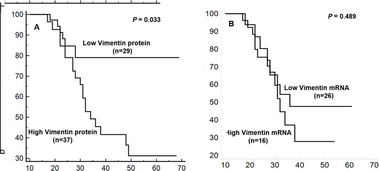
Vimentin protein (p=0.004) and mRNA (p=0.027) expression both showed a significant association with lymph node metastasis. Overexpression of Vimentin protein was observed in 11 of 15 (73%) patients with tumour recurrence and all the four (100%) patients who died of systemic metastasis (table 4). The prognostic significance of Vimentin mRNA and protein expression was determined by Kaplan-Meier analysis. Patients with high Vimentin protein expression showed reduced disease-free survival (p=0.033). However, no significant association was found between Vimentin mRNA expression and disease-free survival (p=0.489) (figure 2).
Kaplan-Meier survival curves showing disease-free survival rates in patients with eyelid sebaceous gland carcinoma according to the status of Vimentin protein and mRNA expression. (A) Patients with high Vimentin protein expression showed worse prognosis than those with low Vimentin protein expression (p=0.033). (B) Patients with high Vimentin mRNA expression did not show any significant association with poor disease-free survival (p=0.489). mRNA, messenger RNA. Titles and legends to supplementary figures.
Univariate analyses of survival
The univariate Cox regression analyses of clinicopathological features related to patient prognosis were performed in all 66 patients with eyelid SGC. Vimentin protein expression (p=0.012) and advanced tumour stage (p=0.002) both emerged as significant risk factors by univariate analysis (table 6). Orbital invasion also showed a trend towards poor disease-free survival (p=0.066) (table 6).
Univariate analysis of risk factors affecting disease-free survival in patients with eyelid sebaceous gland carcinoma (COX proportional-hazard regression model)
Discussion
EMT is an important phenomenon in tumour invasion and metastasis, in which Vimentin acts as a cytoskeletal regulator leading to migration of mesenchymal cells associated with loss of cell adhesion molecule E-cadherin. This is mediated by key transcription factors such as ZEB2, Slug and certain miRNA-200 family members. In our earlier study, we have shown that aberrant expression of EMT markers ZEB2, E-cadherin and miRNA-200 family members (miRNA-200c/141) correlates with poor prognosis of eyelid SGC.21 22 The aim of this study was to determine the expression and prognostic significance of Vimentin in eyelid SGC. Vimentin overexpression has been associated with various malignancies including malignant melanoma, CNS tumours, breast cancer, prostate cancer and gastrointestinal tumours.6
In the present study, cytoplasmic overexpression of Vimentin was observed in 56% of eyelid SGC cases, but it was absent in adjoining normal epithelium. Cytoplasmic localisation of Vimentin has been earlier reported in various cancers including gastric, breast and uterine cervix cancer.23 Interestingly, Luo et al have reported nuclear positivity of Vimentin in nasopharyngeal carcinoma.24 Other studies on glioblastoma multiforme cell lines25 and human neuroblastoma cell lines have also reported nuclear localisation of Vimentin.26 However, currently, very little is known about the molecular mechanisms underlying nuclear translocation of Vimentin. Immunohistochemical findings of our study were further validated by qpCR on 42 fresh frozen tissues. mRNA analysis is useful to identify the molecular mechanisms involved in dysregulation of EMT pathway in tumours. Increased Vimentin mRNA expression was found in 38% cases. The high concordance between Vimentin gene and protein expression further validates the importance of Vimentin as an EMT marker in SGC. Similar results of high Vimentin protein and gene expression have been reported in gastric cancer27 and colorectal cancer.28
Vimentin overexpression correlated with clinicopathological high-risk features of SGC including advanced tumour stage (p=0.002), large tumour size (p=0.023) and pagetoid spread (p=0.05). Correlation of Vimentin expression with advanced clinical stage was also observed in nasopharyngeal24 and colorectal cancer.29 Analogous findings for association of Vimentin expression with higher T group have been reported for non-small-cell lung cancer,16 colorectal cancer28 and pancreatic ductal adenocarcinoma.30
Univariate survival analysis of patient with eyelid SGC cohort showed that high Vimentin immunoexpression was an indicator of adverse outcome. Vimentin also emerged as a predictor of lymph node metastasis, indicating that EMT was present in aggressive SGC cases. Various studies on malignancies such as nasopharyngeal carcinoma,24 colorectal cancer,28 head and neck squamous cell carcinoma31 and oral squamous cell carcinoma32 have demonstrated Vimentin overexpression as predictor of lymph node metastasis and poor prognosis.
There was an inverse correlation between Vimentin and E-cadherin immunoexpression in this study. Vimentin induces a mesenchymal phenotype associated with reduced E-cadherin levels in gastric cancer,27 oral squamous cell carcinoma33 and hepatocellular carcinoma.34 The expression of EMT associated transcription factor ZEB2 showed a direct significant association with Vimentin at both mRNA and protein level, further validating that Vimentin contribute towards EMT in eyelid SGC. Similar findings of association between Vimentin and ZEB2 expression are reported in renal cell carcinoma,14 and invasive epithelial breast cancer cell lines MDA-231, BT549 and Hs578T.35 Overexpression of Slug was observed in only few SGC cases and it did not correlate with Vimentin expression.
To the best of our knowledge, overexpression of Vimentin and its contribution towards EMT in eyelid SGC have been demonstrated for the first time. Its overexpression was related to tumour metastasis and poor survival of patients with SGC and hence can be recommended as a predictor of poor outcome.
References
Footnotes
Contributors SS, KC, AS: Conception and design. MB: Analysis, interpretation and manuscript preparation. SK, NP, MSB, SB: Acquisition of clinical data.
Funding Funding support for the study was provided by All India Institute of Medical Sciences (grant no.: F.8-262/A-262/2014/RS) and Indian Council of Medical Research (grant 3/2/2/240/2014-NCD-III).
Competing interests None declared.
Patient consent for publication Obtained.
Ethics approval The study was conducted after approval from Institute Ethics Committee, AIIMS, New Delhi (Ref. No. IEC/NP-250/03.07.2014) and informed consent was obtained from all patients participating in this study. The study was carried out in accordance with the tenets of the Declaration of Helsinki principles.
Provenance and peer review Not commissioned; externally peer reviewed.
Linked Articles
- At a glance