Article info
- Correspondence:Dr Gurdeep Singh Department of Pediatric Ophthalmology, Aravind Eye Care System, Aravind Eye Hospital and Post Graduate Institute of Ophthalmology, Avinashi Road, Coimbatore- 641 014, Tamilnadu, India; email: gurdeep9999{at}yahoo.co.in
Citation
Publication history
- First published July 17, 2007.
Video Report
Video Report
Pigmented free-floating iris cysts
Gurdeep Singh(1) Veerappan R Saravanan(2) Kalpana Narendran(1) V. Narendran(2)1. Department of Pediatric Ophthalmology, Aravind Eye Hospital and Post Graduate Institute of Ophthalmology, Avinashi Road, Coimbatore- 641 014, Tamil Nadu, India.
2. Retina and vitreous services, Aravind Eye Hospital and Post Graduate Institute of Ophthalmology, Avinashi Road, Coimbatore- 641 014, Tamil Nadu, IndiaCorrespondence: Dr Gurdeep Singh
Email: gurdeep9999{at}yahoo.co.in Department of Pediatric Ophthalmology, Aravind Eye Care System, Aravind Eye Hospital and Post Graduate Institute of Ophthalmology, Avinashi Road, Coimbatore- 641 014, Tamilnadu, India. Tel: +91-0422-4360400; Fax: +91-0422-2593030.Date of acceptance:5th June 2007

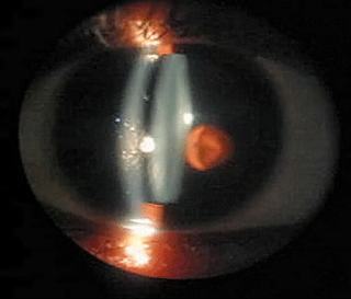
Slit lamp biomicroscopy videos showing the free floating iris cysts in the anterior chamber in case 1 and in the vitreous cavity in case 2. View Video: Fast connectionView Video: Dial up connection
Note: This video is best viewed in Quicktime
Introduction
Free-floating iris cysts are rare. Establishing the exact etiology is hence difficult. These cysts may be located in the vitreous or the anterior chamber. Anterior chamber cysts can be idiopathic or induced by trauma or surgery. Vitreous cysts may be associated with the remnants of the hyaloid system and therefore be congenital, or can result from trauma or ocular disease. We report two patients with free-floating iris cyst, one in the anterior chamber and the other in the vitreous, which was found incidentally on routine eye examination.
Case Report
Case 1: An 8 year old girl presented for routine follow-up with a past history of undergoing traumatic cataract extraction with intraocular lens implantation OD one year prior. Her best-corrected vision was 20/20 in the right eye. Her intraocular pressure was 16mmHg. She had no complaints of any bothersome symptoms. Slit lamp examination showed a free floating dark brown, pigmented round iris cyst in the anterior chamber OD.
Case 2: A 9-year male boy presented for routine eye examination as he was using glasses for refractive error. He had no history of floaters. Anterior segment was unremarkable in the right eye but examination of the anterior vitreous showed a large pigmented cystic lesion. It was free floating and appeared to be composed of pigmented cells joined together by a semi-translucent membrane. Indirect ophthalmoscopy showed no fundus abnormalities, and revealed that the vitreous cyst was probably composed of pigmentary epithelium.
Discussion
Free-floating intraocular cysts can be divided into those occurring in the anterior chamber and the vitreous cavity. According to Clapp, Businelli first described free-floating anterior chamber iris cysts in 1868.1 Free-floating iris cysts in the anterior chamber are a rare, and can be congenital or associated with trauma, surgery, medications or tumors. The wall usually comprises iris pigment epithelium containing pigment granules. The cysts can get detached from the iris surface and float freely in the anterior chamber, or can get reattached to the iris at some other location. They can remain stable or can grow and cause visual disturbance or secondary glaucoma. Visually symptomatic cysts or cysts causing glaucoma need to be removed surgically or collapsed using NdYAG laser.
Tansley was the first to describe free-floating vitreous cysts in 1899. Nork and Millecchia, and Jones have recently summarized their characteristics.2,3 The origin of vitreous cysts has been the source of controversy. It has been proposed that they may be associated with the remnants of the hyaloid system and therefore are congenital, or they can result from trauma, retinal surgery or other ocular disease.3,4 Bilateral cases have been reported in patients with retinitis pigmentosa.3 Orellana and colleagues suggested that the cysts originated from the displacement of pigmented ciliary epithelium from the region of the pars plana, possibly after blunt trauma.5 More recently this conclusion has been questioned by Nork and Millecchia2 who contend that they are congenital and can be regarded as a choristoma of the primary hyaloid system. Mobile, dislodged iris cysts can also be found in the vitreous chamber.6 Rarely, they can lead to visual disturbances if they are imposed on the visual axis. Patients who are significantly symptomatic may elect to receive treatment. Orellana and colleagues removed a vitreous cyst via a pars plana approach, aspirating it with a 20-gauge needle, which allowed the cyst to collapse around the tip of the needle.5 Awan suggested disrupting the cyst with argon laser photocystotomy as a simpler and safer method.7
In our series the patient with the anterior chamber cyst was followed up for a period of two years and the patient with vitreous cyst was followed up for a period of three years without any increase in the size of the cysts or the occurrence of complications.
References
- Clapp CA. Free cyst in the anterior chamber. Am J Ophthalmol 1928; 11: 862-864.
- Nork TM, Millecchia LL. Treatment and histopathology of a congenital vitreous cyst. Ophthalmology 1998; 105: 825-830.
- Jones WL. Free-floating vitreous cyst. Optom Vis Sci 1998; 75: 171-173.
- Asiyo-Vogel MN, el-Hifnawi el-S, Laqua H. Ultrastructural features of a solitary vitreous cyst. Retina 1996; 16: 250-254.
- Orellana J, O’Malley RE, McPherson AR,Font RL. Pigmented free-floating vitreous cysts in two young adults. Ophthalmology 1985; 92: 297-302.
- Shields JA, Kline MW, Augsburger JJ. Primary iris cysts: a review of the literature and report of 62 cases. Brit J Ophthalmol 1984; 68: 152-166.
- Awan KJ. Biomicroscopy and argon laser photocystotomy of free-floating vitreous cysts. Ophthalmology 1985; 92: 1710-1711.
Files in this Data Supplement:
Request permissions
If you wish to reuse any or all of this article please use the link below which will take you to the Copyright Clearance Center’s RightsLink service. You will be able to get a quick price and instant permission to reuse the content in many different ways.


