Article Text

Abstract
Aim: To determine the efficacy of meloxicam ophthalmic formulation on COX-2 activity and expression, inflammation-related cytokines expression and inflammation in an ocular inflammation model.
Methods: Ocular inflammation was induced in New Zealand rabbits by topical application of croton oil (3%) for 3 h. An ophthalmic solution of 0.03% meloxicam, 0.1% sodium diclofenac or vehicle (Sophisen™) was administered every 4 h. Conjunctiva, cornea, aqueous humour and vitreous humour were collected.
Results: In irritated eyes, 72 h of meloxicam treatment downregulated COX-2 expression and activity (mRNA by RT-PCR and PGE2 levels by ELISA, respectively) in a time-dependent manner and reduced inflammation. Meanwhile, diclofenac failed to reduce COX-2 mRNA or PGE2 to basal levels after 7 days of treatment. Meloxicam treatment downregulated IL-6 and IFN-γ expression in the conjunctiva and IL-1β and TNF-α expression in the cornea. Diclofenac failed to modify these cytokines in both tissues. Meloxicam treatment increased the expression of IL-6 in conjunctiva, and IL-10 in cornea, while diclofenac had no effect on these cytokines.
Conclusion: Meloxicam treatment was more efficient than diclofenac in downregulating the expression and activity of COX-2, reducing inflammation, and modifying the inflammatory-related cytokines.
Statistics from Altmetric.com
Ocular irritation involves the release of pro-inflammatory cytokine interleukin (IL)-1 that induces the expression of other cytokines, such as IL-6 and IL-8.1 In turn, chemokines recruit leucocytes to the eye and promote the synthesis of inflammatory mediators such as prostaglandin E2 (PGE2). Expression of cyclooxygenase 2 (COX-2), is induced by inflammation, and COX-2 has been principally involved in the generation of PGE2 in several inflammatory models.2
Non-steroids anti-inflammatory drugs (NSAIDs) such as aspirin suppress the nuclear transcription factor κB (NFκB) which controls the expression of COX-2.3 Increased COX-2 activity upregulates several components of the signalling cascade of activation of some cytokines and is intimately related with the inflammatory process.4 IL-1β secretion induces PGE2 production through activation of COX-2 and cJun-N-terminal kinase.5 Other known regulators of COX-2 expression are TNF-α, TGF-β1,6 and IL-6.7 IL-6 is a pleiotropic cytokine that influences inflammatory reactions.8 TNF-α has been implicated in cutaneous inflammation involving keratinocytes.9
Meloxicam is a new preferential COX-2 inhibitor currently used for the treatment of osteoarthritis and rheumatoid arthritis as a systemic drug. Due to the marked selectivity of meloxicam over diclofenac for COX-2 inactivation,10 it has been proposed that meloxicam can be used as a topical ophthalmological drug to reduce inflammation without altering the normal housekeeping functions of COX-1 and other PGs.
MATERIALS AND METHODS
Animals and samples
Male New Zealand rabbits weighing 1.9–2.2 kg were housed on a 12-h light, 12-h dark schedule and allowed free access to food and water. Animals were killed by CO2 inhalation, and the conjunctiva, cornea, vitreous humour, and aqueous humour were dissected and immediately frozen in liquid nitrogen. The Mexican Regulatory Guideline for the Use of Animals in Research (NOM-062-ZOO-1999) was followed.
Croton oil-induced ocular inflammation and anti-inflammatory treatment
Rabbit ocular inflammation was induced by applying a topical solution of 3% croton oil in 2-ethoxyethanol, as previously described.11 Three hours after croton oil-induced inflammation, right eyes were treated every 4 h for a duration of 7 days with 0.03% meloxicam formulation, 0.1% diclofenac formulation or vehicle (Sophisen, patent number 6071958, USPTO). Different endpoints were evaluated 0, 4, 24, 72 and 144 h after croton oil treatment (four animals per group). Rabbits were killed and tissues processed for mRNA and PGE2 quantification.
Quantification of mRNA by semiquantitative RT-PCR
Total RNA was extracted using Trizol® reagent according to the manufacturer’s recommendations and quantified by UV photometry as previously reported.12 Specific mRNA levels were determined by semiquantitative RT-PCR using 18S rRNA as an internal control (all chemicals were Invitrogen). Briefly, 1 μg of RNA was incubated with 250 ng of random primers, and 200 U of reverse transcriptase (SuperScript III) for 30 min at 50°C. A 1:10 dilution of cDNA was subjected to PCR amplification using 20 pmol of primers and 0.5 U of Platinum Taq DNA polymerase. The cycling programme was: 3 min incubation at 94°C, 12–47 cycles of amplification, denaturation for 50 s at 94°C, primer annealing for 40 s (table 1), extension for 40 s at 72°C, and 7 min extension at 72°C. Ten microlitres of the PCR products was separated by electrophoresis on a 2% agarose gel. Gels were stained with ethidium bromide, and PCR products were quantified by densitometry with the Image J software (http://rsb.info.nih.gov/ij/). RT-PCR products were normalised per amount of 18S rRNA product.
PGE2 enzyme-linked immunosorbent assay (ELISA)
PGE2 levels were analysed using the prostaglandin E2 Biotrak ELISA system (Amersham Biosciences, Piscataway, NJ) according to the manufacturer’s instructions.
Histophatology
In a different group of animals, treatment with meloxicam or diclofenac ophthalmic formulations were applied chronically every 4 h for 3 weeks, and eyes were collected and cryopreserved. Tissues were processed for further histopathological analysis as previously reported,13 slides were stained with haematoxylin–eosin and microscopically analysed.
Statistical analyses
Data are presented as the mean (SD). Statistical significance was assessed using a one-way ANOVA, and Student t test; differences were considered significant when p<0.05.
RESULTS
Basal COX-2 mRNA levels were higher in the cornea than in conjunctiva (fig 1, p<0.05). However, compared with basal COX-2 mRNA levels, croton oil application induced a threefold increase in COX-2 mRNA levels in the conjunctiva and only a twofold increase in the cornea 3 h after application (fig 1). Without croton oil treatment, PGE2 levels were barely detectable in the aqueous humour; however, croton oil induces a strong increase in PGE2 compared with basal levels (table 2). Vitreous humour had higher PGE2 levels than aqueous humour, but its levels did not change with croton oil application (459 (29), and 21 (14) pg/mL, respectively). Croton oil also failed to modify basal PGE2 levels detected in the cornea and conjunctiva.

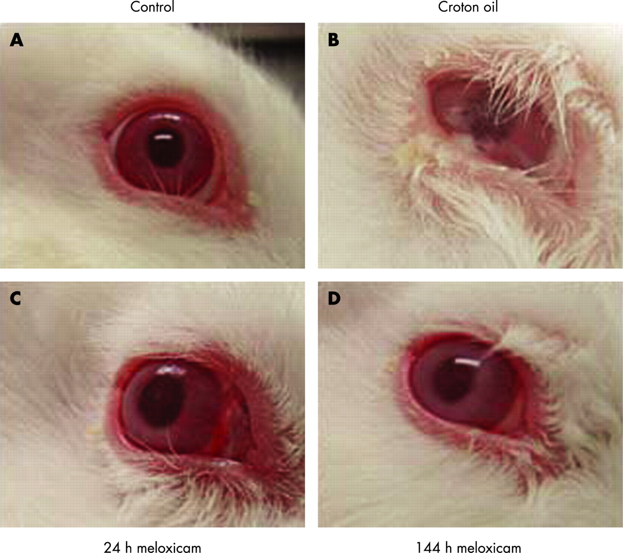
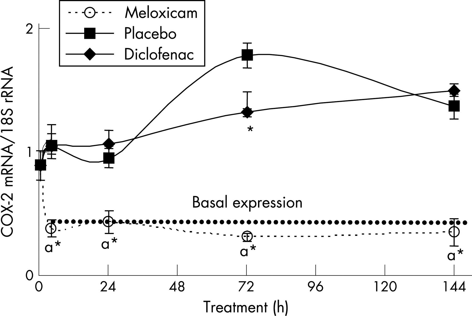
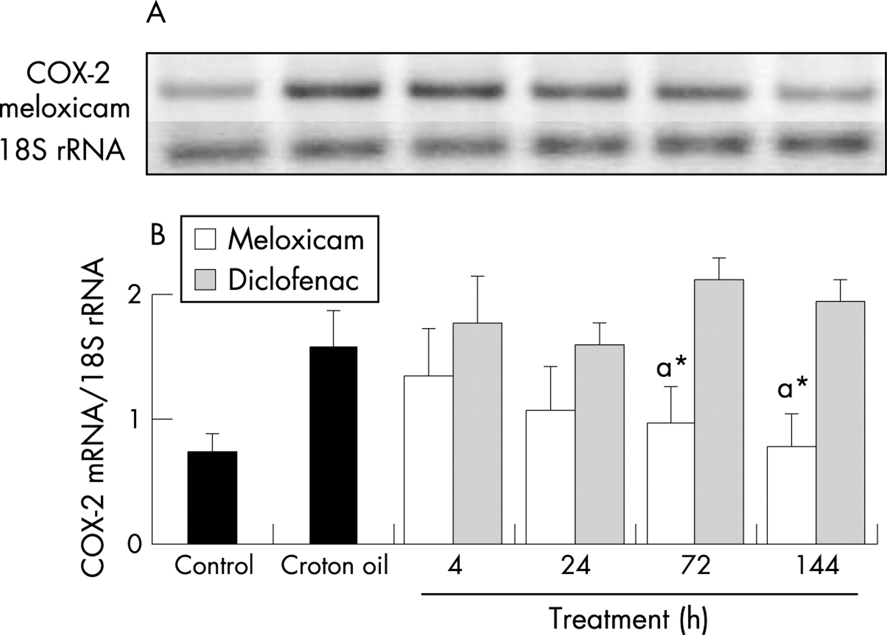
Ophthalmic solution with 0.03% of meloxicam efficiently downregulated COX-2 mRNA expression in conjunctiva to basal levels after 4 h of treatment (fig 2). The ophthalmic formulation containing 0.1% of diclofenac failed to downregulate COX-2 mRNA expression back to basal levels through 144 h of treatment but did reduce COX-2 mRNA levels by 26% at 72 h of treatment compared with placebo. As shown in table 2, croton oil application induced a strong increase in PGE2 concentrations, and the placebo-treated animals maintained high levels of PGE2 that decreased only after 144 h, which is consistent with the normal course of ocular inflammation observed in rabbits (not shown). Treatment with diclofenac induced a rapid, but transient, reduction in PGE2 levels that increased again dramatically at 24 h in aqueous humour (table 2). Meanwhile, treatment with meloxicam induced a gradual, but steady, reduction in PGE2 concentration. Compared with croton oil treatment alone, meloxicam downregulated COX-2 mRNA levels in the cornea in a time-dependent fashion, while diclofenac treatment did not modify COX-2 expression (fig 3). All the groups of animals treated with croton oil showed the same clinical signs of inflammations, such as swelling, redness, and ocular secretion, the only difference being that meloxicam treatment reduced these clinical signs after 72 h of treatment (p<0.05, fig 4), while diclofenac treatment yielded the same results only after 144 h of treatment as did the placebo (not shown). After 144 h, all the animals had the same appearance as the control animals, and no other signs were observed or were different between groups. Regardless of treatment, by 144 h all eyes had returned to the basal condition (fig 4).


The basal profiles of inflammatory-related cytokines expressed were different between the conjunctiva and cornea. In conjunctiva, IL-1β, IFN-γ, TNF-α, IL-6 and IL-10 were all expressed prior to induction of ocular inflammation (fig 5). Also, croton oil induced a small but significant increase in the pro-inflammatory cytokines, IL-1β, IFN-γ, and TNF-α, and a strong increase in IL-6 (fig 5, p<0.05). However, there was no upregulation of IL-10 (data not shown). Meloxicam treatment did not change the levels of IL-10; nor did treatment with diclofenac (data not shown). Meloxicam significantly increased IL-6 mRNA levels, while diclofenac failed to modify IL-6 levels (fig 5B). Treatment with meloxicam significantly reduced IL-1β mRNA level at 72 and 144 h, while diclofenac treatment had no effect on IL-1β levels (fig 5C). Compared with croton oil induction, treatment with meloxicam decreased IFN-γ mRNA levels at 72 h, while diclofenac, again, did not change the expression of IFN-γ (fig 5D). Meloxicam also significantly reduced TNF-α expression to the basal level from 24 h of treatment, while we observed an increase on TNF-α expression with diclofenac treatment that was significant at 72 and 144 h (fig 5E).

In cornea, there was no basal expression of any of the cytokines evaluated before induction of ocular inflammation (fig 6). Levels of TNF-α, IL-10 and IL-1β were significantly upregulated by croton oil treatment (fig 6, p<0.05), but upregulation of IFN-γ or IL-6 was not detected in this tissue (data not shown). IL-10 mRNA levels were increased with meloxicam treatment from 24 h, while diclofenac did not modify IL-10 expression (fig 6B). Meloxicam reduced the expression of IL-1β at 72 and 144 h of treatment, and diclofenac failed to modulate IL-1β expression (fig 6C). Meloxicam rapidly decreased TNF-α to basal levels (0 pg/ml) by 4 h treatment and maintained this through 144 h treatment (fig 6D); in comparison, diclofenac treatment only reduced TNF-α expression at 4 h, and it was unchanged thereafter.

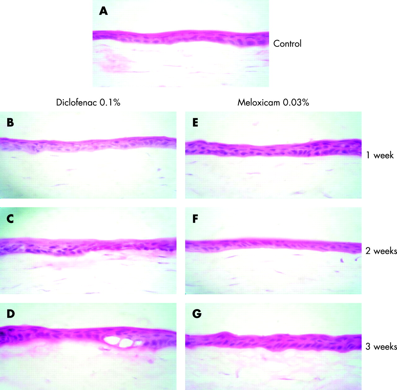
Finally, despite all the changes on cytokines induced by diclofenac, or meloxicam treatments, we observed no physical changes in the structure of the cornea even after 3 weeks of chronic application of meloxicam (fig 7).
DISCUSSION
COX-2 is a highly inducible enzyme that regulates diverse actions on various cell types and functions.14 Enhanced COX-2 expression may be harmful, inducing the production of atheromatous plaque instability and hyperplasia.15 The induction of COX-2 transcriptional activation by pro-inflammatory mediators and cytokines has been extensively characterised,16 and we observed that meloxicam; a selective inhibitor of COX-2 activity,10 exerts a clear downregulatory effect on COX-2 gene expression. This effect is not easily explained, since it has not been described previously that an enzymatic substrate of COX-2 could regulate gene expression. Although several stimuli are known to regulate COX-2 expression, the molecular mechanisms by which cytokines can modulate COX-2 expression are poorly understood. On the one hand, IFNγ-priming (50–500 U/ml) downregulates COX-2 expression in a time- and dose-dependent fashion. Furthermore, IFN-γ is able to inhibit COX-2 gene expression in response to IL-1β but fails to do so under a different stimulus, such as LPS.17 Expression of IL-6 and COX-2 genes in the cell seems to be differentially regulated by cytokines through prostaglandin-dependent and -independent pathways.18 The COX-2 DNA sequence contains several potential transcription regulatory sequences, including a TATA box, a C/EBP motif, two AP-2 sites, three SP1 sites, two NF-κB sites, a CRE motif, an Ets-1 site,19 p300 and CREB binding sites,20 and nuclear factor of activated T cells binding site (NFAT).21 Although many cytokines have been described to modulate their own gene transcription or transcription of other genes by a crosstalk or interacting with these transcriptional factors, none of them is related to the phenomena of direct inhibition of mRNA expression by an enzymatic substrate or by-product of enzymatic activity. Thus, new research concerning the mechanisms involving enzymatic activity and gene expression regulation is needed and would probably be the key for the regulation of gene expression as a therapeutic measure, not only in inflammatory processes, but in many other diseases directly related with the modulation of particular genes expression. Although the modulation of proinflammatory cytokines does not seem to completely explain the efficacy of meloxicam in downregulating the ocular inflammation, at least we can say that the best biomarkers used in this case are the PGE2 release (as marker of COX-2 activity), together with IL-1β (as a marker of inflammation). Regardless of the amount of proliferative cytokines induced by the treatment with meloxicam, these cytokine levels did not alter the corneal tissue, at least at the structural level, even after 3 weeks of chronic treatment. Although it is well known that some inflammatory-related cytokines have proliferative actions over normal keratinocytes, our results demonstrated that the regulation on the expression of cytokines by meloxicam is not directly related to the maintenance and integrity of the corneal tissue at this level.
In conclusion, treatment of ophthalmic inflammation with a solution of 0.03% meloxicam reduced COX-2-related activity and inflammation-related cytokine release more effectively than a 0.1% diclofenac ophthalmic formulation. Meloxicam also presents some additional advantages, as it is a more specific COX-2 inhibitor than diclofenac10 and thus may produce fewer secondary effects due to the inhibition of the constitutive COX-1 enzyme.
Acknowledgments
The authors thank Dr Jorge Fernández Hernández from Cinvestav for his technical assistance.
REFERENCES
Footnotes
Funding: This project was financed by Laboratorios SOPHIA SA de CV, Mexico.
Competing interests: None declared.