Article Text

Abstract
Aim: To examine the acute effects of wearing swimming goggles upon intraocular pressure (IOP).
Methods: This research consisted of a Pilot study and a Validation study. Holes were drilled into the faces of 13 different goggles to allow IOP measurement by applanation tonometry. IOP was measured before goggle wear, 2 min after goggle application, 20 min after goggle application and after goggle removal. The Pilot study (n = 15) was initially performed to investigate changes in IOP while wearing five different types of swimming goggles. Anatomical and goggle design parameters from the Pilot study were then used to generate a predictive model and design a Validation study (n = 20). The Validation study tested the predictive model, examined IOP changes using another eight goggles and clarified whether IOP changes were sustained for the duration of goggle wear.
Results: IOP increased while wearing goggles by a mean pressure of 4.5 mm Hg (SD 3.7, p<0.001) with this pressure rise being sustained for the duration of goggle wear. A smaller goggle face area (p = 0.013), was consistently associated with greater IOP elevation.
Conclusion: These measurements were not taken while swimming, but they suggest that some swimming goggles can elevate IOP.
Statistics from Altmetric.com
Swimming is a popular form of exercise with many swimmers wearing goggles to improve underwater visibility. Tension from the goggle headband keeps the goggles in place. This force acting on the goggles may compress orbital vasculature and other structures to cause an elevation in intraocular pressure (IOP). Continuously elevated IOP is a significant risk factor for glaucoma development and progression.1 2
There is no previous information regarding the effects of swimming goggles upon IOP. There are case reports of migraine,3 supraorbital neuralgia,4 eyelid swelling,5 skin irritation6 and diplopia7 associated with wearing goggles. One study found that the air pressure between a swimming goggle and the eye decreased as one subject placed his goggle on and off.8 We wanted to determine if goggle wear resulted in immediate changes to IOP and if these changes were sustained for the duration of goggle wear. We were also interested in determining the goggle characteristics that were associated with any IOP changes. We performed a Pilot study to test the immediate effect of wearing goggles upon IOP and used measurements of the subjects’ orbits and swimming goggles to generate a predictive model of IOP change. A subsequent Validation study involving more subjects and using a greater range of goggles tested the validity of this predictive model. The Validation study also added to the data from the Pilot study and allowed us clarify if IOP changes upon goggle application were sustained or varied while wearing goggles for an extended period of time.
MATERIALS AND METHODS
This research consisted of an initial Pilot study and a subsequent Validation study. The study was approved by the University of Western Australia Human Ethics Committee and was performed in accordance with the Declaration of Helsinki. Informed consent was gained from all subjects or their parents.
Swimming goggle characteristics and measurements
All goggles used for this study were designated a number from 1 to 13. Numbers 1 to 5 were used for the Pilot study, and numbers 6 to 13 were used for the Validation study. The Pilot study was designed to test a small but broad sample of swimming goggles using a protocol that allowed the collection of a large number of IOP measurements (table 1, goggles 1 to 5). For the Validation study, we used eight different goggles (table 2, goggles 6 to 13), representing the broadest range of goggles commercially available in Perth.
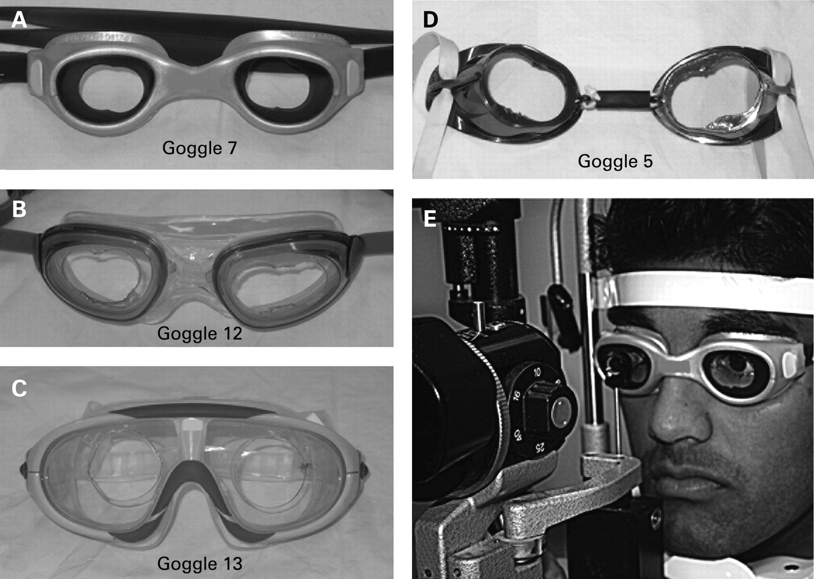
The majority of goggles consisted of two separated rigid plastic eye cups with a rubber cushioning seal surrounding the lip of each cup. Exceptions were goggle number 5, which had no rubber seal, and goggle numbers 12 and 13, which had a larger single eye piece which covered both eyes and had air continuity between the eyes (fig 1). One 2 cm diameter hole was drilled into the front of each goggle eye piece so that applanation tonometry could be performed through the hole.

Several parameters were measured for each goggle (fig 2). Vertical and horizontal goggle widths were measured across the internal aspect of each eyepiece from rubber seal to rubber seal. The goggle face area was defined as the area of skin enclosed by each eyepiece and was assumed to be elliptical in shape. The formula to calculate the area of an ellipse [vertical width×horizontal width×π/4] was used to calculate the goggle face area. The horizontal-to-vertical goggle width ratio was also calculated. Goggle number 5 had no rubber seal, and the diameter for this goggle was measured across the internal aspect of the rigid plastic cup. Goggles 12 and 13 had no contact points over the nasal bridge, so we measured horizontal width to the midpoint. The depth of the rubber seal was measured at four points evenly distributed around each goggle, and the width of the rubber seal was measured at eight positions around the face of all goggles. The mean values of these measurements were used for analysis.

Orbital measurements
The vertical and horizontal orbital margin widths were measured using Vernier callipers, taking the measurement across the pupil with each subject looking in the primary position. The orbital area was calculated as vertical width×horizontal width×π/4. The horizontal-to-vertical orbital width ratio was also calculated. The age, sex and central corneal thickness (Quantel, Pocket Pach 2, Clemont, France) was also recorded for each subject.
Determination of baseline IOP
The aim of this study was to measure the additive effect of goggle wearing upon IOP. Tonographic studies have shown that increased IOP induced by an applied force leads to increased aqueous egress from the eye and a transient decrease in IOP when the force is removed.9 To account for any tonographic effect as a result of swimming goggle wear, we measured IOP prior to and after goggle wearing, then used the mean of these two measures as the baseline IOP. The change in IOP as a result of swimming goggle wear was calculated with respect to this baseline IOP. We also analysed the difference between the IOP recorded before goggle application and after goggle removal.
Pilot study
For the Pilot study, 15 subjects were recruited from a local swimming club. Following installation of 0.5% proxymetacaine and fluorescein eye-drops, the IOP from each eye was measured using Goldmann applanation tonometry before swimming goggles were worn. Subjects were instructed to wear each goggle in a random sequence and adjust them as they would when they entered the swimming pool. Two minutes after they had adjusted their goggles and were comfortable, the IOP was measured (fig 1E). Those goggles were then removed and another set worn with the IOP measured after 2 min. This sequence was repeated until each of the five goggles had been tested. Five minutes after the fifth goggle had been removed, IOP was recorded with no goggles being worn. Fourteen subjects were then instructed to put on and wear either a small (no. 1) or large (no. 4) width goggle for 20 min with the IOP measured at this point. Five minutes following removal of the goggles, the IOP was again measured.
Validation study
The Validation study involved testing eight other goggles upon 20 new subjects. The protocol was similar to the Pilot study with the following differences. Each subject wore two or three sets of goggles, with the third set being optional depending upon the time commitments of the subject. IOP measurements were taken prior to goggle application, after 2 min of goggle wear and after 20 min of wear. The goggles were then removed, and 5 min later another IOP measurement was recorded.
The subject then applied the second or third goggle set, and the same IOP measurement protocol was used.
Statistical analysis
Only right eye data were used, and the mean and standard deviations of measurements are presented. All calculations were performed using SigmaStat (Systat Inc, Point Richmond, CA). A paired Student t test before and after intervention, reporting 95% confidence intervals (CI), was used. Using the 2 min Pilot study data, a multiple linear regression model was used to investigate the significant variables that were associated with a change in IOP. The dependent variable, IOP change, was modelled against sex, age, goggle face area, horizontal-to-vertical goggle width ratio, mean rubber width, mean rubber depth, central corneal thickness, orbital area, horizontal-to-vertical orbital width ratio and baseline IOP. Sex was scored as a binary variable (male 1, female 0). The model was calculated repeatedly with removal of the least significant variable until only significant variables remained (p<0.05). Once the predictive model was determined, we used goggle and anatomical data from the Validation study to predict the rise in IOP. Because we were interested in predicting IOP rise and were extrapolating beyond the range of goggle parameters used in the Pilot study, we converted all negative IOP change predictions to zero. The Pearson least mean squares correlation coefficient was then used to report the correlation between the predicted and actual IOP rise.
RESULTS
Demographic, anatomical and swimming goggle measurements
Fifteen subjects were examined for the Pilot study with a mean age of 22.7 (SD 15.9) years. All subjects were Caucasian, eight males and seven females. For the Pilot study, a total of 88 right eye IOP measurements were taken, being 74 measurements at 2 min after goggle application and 14 measurements after 20 min application (table 1). One subject would not allow measurement of IOP from the right eye. The mean baseline IOP was 14.9 (3.9) mm Hg with a mean central corneal thickness of 536 (32) µm. The mean vertical orbital width was 28.7 (2.6) mm (range 24.5 to 32.5), and the mean horizontal orbital width was 37.9 (3.4) mm (range 31 to 43). There was no significant difference in vertical (p = 0.98) or horizontal (p = 0.56) orbital widths between males and females in this group.
Twenty subjects (10 males and 10 females) were examined in the Validation study with a mean age of 35.6 (20.4) years. There was a difference in racial mix in this group, with two subjects from China, three from the Indian subcontinent and the remainder being Caucasian. A total of 43 right eye IOP measurements were taken from the 20 subjects at each time point (two measurements from 17 subjects and three measurements from three subjects; table 2). The mean baseline IOP (13.5 mm Hg, p = 0.217), age (p = 0.053), central corneal thickness (p = 0.154), vertical (p = 0.295) and horizontal (p = 0.244) orbital widths were not significantly different between the two study groups.
IOP changes following swimming goggle wear
All goggles with the exception of goggle 13 caused significant IOP elevation, with goggles 1 and 9 causing a mean 10.1 mm Hg (p<0.001) and 13.4 mm Hg (p<0.005) elevation respectively after 20 min of goggle wear. The mean IOP rise from all goggles was 4.5 mm Hg (SD 3.7, p<0.001). The key physical properties of each pair of goggles used in the Pilot study and Validation study and the mean IOP rise induced by each of these goggles are described in tables 1, 2 respectively.
In the Validation study, the mean difference in IOP between measurements taken 2 min after goggle wear and 20 min after goggle wear was an insignificant 0.023 mm Hg (p = 0.91, df = 42; table 2). The IOP measurement taken 5 min after removal of the first goggle decreased by a mean of 1.75 mm Hg with respect to the IOP measurement taken before goggle application (p<0.001, df = 19). The difference in IOP measurement obtained 5 min after first goggle removal and 5 min after second goggle removal was insignificant (mean drop 0.33 mm Hg, p = 0.513).
Multiple linear regression analysis and the predictive model
Application of a multiple linear regression model to Pilot study data revealed that age, baseline IOP, central corneal thickness, and mean rubber depth, were not significantly associated with IOP rise. The final linear model (r2 = 0.524) revealed that reduced goggle area, (p<0.001), reduced orbital area (p = 0.021), reduced horizontal-to-vertical orbital width ratio (p = 0.04) and reduced rubber seal width (p<0.001) induced a significant rise in IOP. Sex was also significant, with males generally experiencing less IOP rise (p = 0.001).
Using the linear regression results from the Pilot study, we were able to generate the following predictive formula for IOP change: Predicted IOP change = 68.932–(3.294×sex)–(2.009×mean rubber width)–(0.00822×orbit area)–(0.0243×goggle area)–(12.731×horizontal-to-vertical orbit ratio). Using all the Validation study anatomical and goggle measurements, we calculated a predicted IOP change for each subject/goggle combination. The Pearson least mean square regression between the predicted IOP change and the actual IOP change measured was positive (r2 = 0.194). When we applied a multiple regression model to Validation study data to determine the influence of various anatomical and swimming goggle parameters on IOP rise, we found that orbital area (p = 0.773), mean rubber width (p = 0.497), sex (p = 0.309) and horizontal-to-vertical orbital width ratio (p = 0.134) were not significantly associated with IOP change. We found that goggle area was the only variable that was significantly associated (p = 0.013) with change in IOP. Figure 3 plots the change in IOP against goggle face area (r2 = 0.207, n = 56, p<0.001). Data from both Pilot and Validation studies after 20 min of swimming goggle wear are included in this figure.

DISCUSSION
Our experiments, were not performed while swimming, but they demonstrate that goggles may elevate IOP significantly. These results have important implications as an elevation of IOP is known to be a potent risk factor for the causation and progression of glaucoma.2 10
There was no significant difference between the IOPs taken 2 min and 20 min after goggle application, implying that IOP is elevated rapidly and is sustained with goggle wear. We drilled holes in the goggle face to permit IOP measurement which resulted in the loss of an airtight seal and eliminated the usual suction effect. This suction is 0 to −5 mm Hg during comfortable goggle wear.8 This effect is equivalent to moving up 60 m in altitude and would not be expected to influence IOP significantly. Our measurements also do not include the effect of external water pressure upon goggles which causes a 0.74 mm Hg pressure increase per centimetre water depth. This effect increases external pressure on the goggles and would most likely increase the pressure upon the eye.
A smaller goggle size was the most significant and only factor consistently associated with IOP elevation. It is likely that headband tension transmitted into the orbit through the rubber seal increases orbital tissue pressure, compressing the globe and leading to the sustained IOP rise observed. It is also likely that there are anatomical or other factors which interact with goggle size to determine the IOP rise, but we were unable to determine these with our measurements. To date, there have been no studies examining a relationship between wearing goggles and glaucoma, but it appears advisable to warn glaucoma patients about the potential risk of raised IOP while wearing small swimming goggles.
Acknowledgments
Thanks to the West Coast Swimming Club for organising subjects and visits, and SK Morgan for organizing the goggle application, photographs and collating measurement data.
Footnotes
Funding: The McCusker Glaucoma Centre, and National Health and Medical Research Council programme grant 211901 provided financial support.
Competing interests: None.
Ethics approval: The study was approved by the University of Western Australia Human Ethics Committee and was performed in accordance with the Declaration of Helsinki.
Patient consent: Obtained.