Article Text

Abstract
Background: Double-blinded, randomised, prospective, pilot-study to determine the effect of systemic bevacizumab therapy.
Methods: Subjects with fibrovascular pigment epithelial detachment, subfoveal choroidal neovascularisation extending under the geometric centre of the foveal avascular zone and/or macular thickness of at least 300 μm in both eyes were included. Sixteen eyes were included and randomised equally to receive either three infusions of 5 mg/kg Avastin or 100 ml of 0.9% sodium chloride every 2 weeks. The main outcome measure was the lesion size. The follow-up time was 24 weeks.
Results: Throughout the 24-week follow-up, the lesion size and macular thickness decreased in the Avastin group by 0.5 (SD 0.08) mm and 103.6 (14.9) μl respectively. In both groups, visual acuity remained stable in seven eyes and decreased in one eye. At the end of follow-up, 50% of the eyes in the Avastin group became fibrotic, 37.5% remained unchanged, and 12.5% developed a subretinal bleeding. There was a treatable rise in blood pressure after Avastin treatment.
Conclusion: Systemic Avastin could be offered to patients with exudative age-related macular degeneration in both eyes and/or patients who refuse intravitreal injections if blood pressure is normal and there is no history of thrombosis.
Statistics from Altmetric.com
Choroidal neovascularisation (CNV) in age-related macular degeneration (AMD) is one of the major causes of severe vision loss in older people in North America and Europe.1 AMD threatens distance as well as reading acuity, and it is already known that the vascular endothelial growth factor (VEGF) plays a major role in the development of CNV.2
When we began the present study, in June 2005, promising results were published of phase I/II clinical trials after intravitreal injections of the anti-VEGF drugs pegaptanib (Macugen, Eyetech Pharmaceuticals, New York),3 4 ranibizumab (Lucentis, Genentech, South San Francisco, California),5 and systemic treatment with bevacizumab (Avastin, Genentech).6 At that time a systemic treatment, which would need to be administered only a few times within a short time, was very tempting, especially for patients with a low quality of life due to bad visual acuity.
Photodynamic therapy (PDT),7–9 in combination with intravitreal injection of triamcinolone acetonide,10 11 and subretinal surgery12–14 were the common treatments for AMD. These methods either were very invasive or had to be repeated several times over long periods of time in order to show some effect. PDT was performed only in eyes with predominantly classic lesions and pigment epithelium detachments (PED) with a maximum lesion of 50%, and there was little chance of vision improvement in these eyes. There was no treatment available for larger minimally classic and occult-only lesions or lesions including larger PED.6
Bevacizumab was a new anti-VEGF which showed promising results as a combination therapy with 5-fluorouracil, leucovorin and oxaliplatin in first-line treatment of metastatic colorectal cancer15 16 and as systemic therapy against AMD.6 However, only a case series for AMD treatment existed. The rationale of the present study was to determine the effect of systemic bevacizumab therapy in subjects with fibrovascular PED, involving the geometric centre of the foveal avascular zone, in comparison with placebo treatment with sodium chloride 0.9%. The main outcome measure was the lesion size, while best-corrected visual acuity (BCVA), reading visual acuity (RVA) and macular thickness were additional outcome measures. We believed that a decrease in density of the subfoveal CNV may well lead to a decrease in macular thickness, thereby allowing a better BCVA. Systemic Avastin was a promising treatment option for AMD, although it is mainly administered off-label as an intravitreal injection these days.17–23
MATERIALS AND METHODS
The BEAT-AMD-Study is a prospective, double-blinded, randomised, monocentric pilot study, and was conducted at our clinic. The study and data accumulation were carried out with approval from the ethics committee of the city of Vienna, and informed consent for research was obtained from all subjects.
Eight subjects with fibrovascular PED, subfoveal CNV extending under the geometric centre of the foveal avascular zone and/or a macular thickness of at least 300 μm in both eyes were included. Exclusion criteria were proteinuria or renal impairment, hepatic dysfunction, vision-threatening ophthalmic diseases other than AMD or a history of arterial thromboembolic diseases or cancer.
Examinations were performed before the start of infusion therapy, on a weekly basis for the first 6 weeks, every second week for the following 6 weeks, then every 4 weeks for the next 12 weeks. The follow-up time was 24 weeks.
Measurement of BCVA, RVA, intraocular pressure (IOP), macular thickness and complete slit-lamp examination were performed at every visit, while fluorescein angiography (FA) and Indocyanine Green angiography (ICGA) were performed before infusion therapy and after 12 and 24 weeks.
BCVA was determined using the ETDRS charts according to the guidelines of the Treatment of Age-related Macular Degeneration with Photodynamic Therapy (TAP) study.7 It was performed at a test distance of 4 m, with letter-by-letter scoring and a chart luminance of 100 candelas (cd)/m2. A loss or gain of up to two lines (10 letters) was considered as stable BCVA, and a loss of three lines (15 letters) was considered as a decrease in BCVA.
RVA was examined using a standardised, German-language reading test (Radner reading charts).24 The measurements were performed monocularly with the patient’s optimal refractive correction at working distance.
The macular thickness was measured using the Stratus OCT 3 (Carl Zeiss Meditec, Dublin, California). Using the macular thickness program, six radial lines through the centre of the foveal avascular zone were made. An internal fixation light was used to maintain the fixation throughout the examination, where possible.
FA was performed with the Heidelberg Retina Angiograph (Heidelberg Engineering, Heidelberg, Germany). Six to eight photographs were taken in the early phase after injection of fluorescein, then every 15 s, after 1 min, 2, 5 and 10 min. Distances in the angiograms were measured in millimetres with the help of the Heidelberger software. ICGA was also performed, including late frames after 20–30 min.
Subjects were looked after by an internal medical doctor (oncologist) throughout the entire study. Internal medical examinations including blood pressure, blood sampling, urine sampling, electrocardiogram and weight measurement were performed throughout the first 6 weeks and after 24 weeks, in order to avoid any additional risk related to the administration of Avastin.
Subjects were randomised to receive either bevacizumab (Avastin, Genentech) intravenous infusions, three times 5 mg/kg every 2 weeks (Avastin group) or sodium chloride 0.9% intravenous infusions, three times 100 ml every 2 weeks (NaCl group). Randomisation was performed by the oncologist, by picking an envelope. As the present study is double-blinded, the oncologist and the pharmacist, who prepared the infusion, were the only ones informed about the treatment group in which the subject was randomised. The subject and the examining ophthalmologists were not informed about the randomisation result.
Data were collected and calculated using SPSS (Version 11.0). Statistical analysis was not performed due to the small number of subjects included. Data are presented as mean (SD), percentage and difference between measurements at different time points.
RESULTS
Four subjects received Avastin systemic therapy, and four subjects received NaCl infusions. Four women and four men were included. Two women and two men received Avastin, and the others received NaCl. The mean age was 79 (SD 4) (73–82 years) in the Avastin group and 74 (9) (65–83 years) in the NaCl group. All eyes were phakic.
Before treatment, the BCVA in the Avastin and in the NaCl group was 0.33 (0.4) and 0.56 (0.33), respectively, while the RVA was 0.6 (0.5)logRAD and 0.5 (0.4)logRAD respectively. At that time the macular thickness and the lesion size were 375.9 (76.6) μm and 5.42 (2.15) mm respectively in the Avastin group (figs 1, 2) and 263.2 (81.2) μm and 3.72 (1.88) mm in the NaCl group (figs 3, 4). The mean blood pressure was 140/80 mm Hg in both groups. The IOP was 16 (1) mm Hg in the Avastin group and 16 (4) mm Hg in the NaCl group.

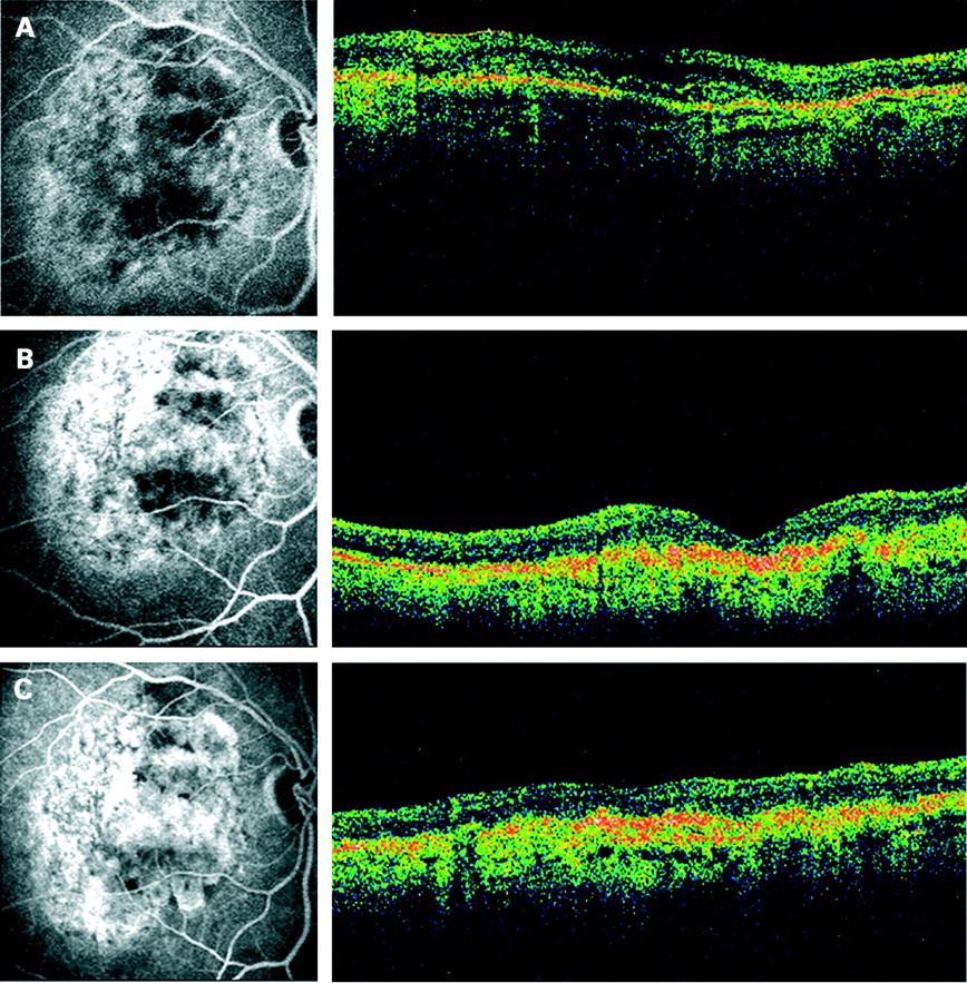
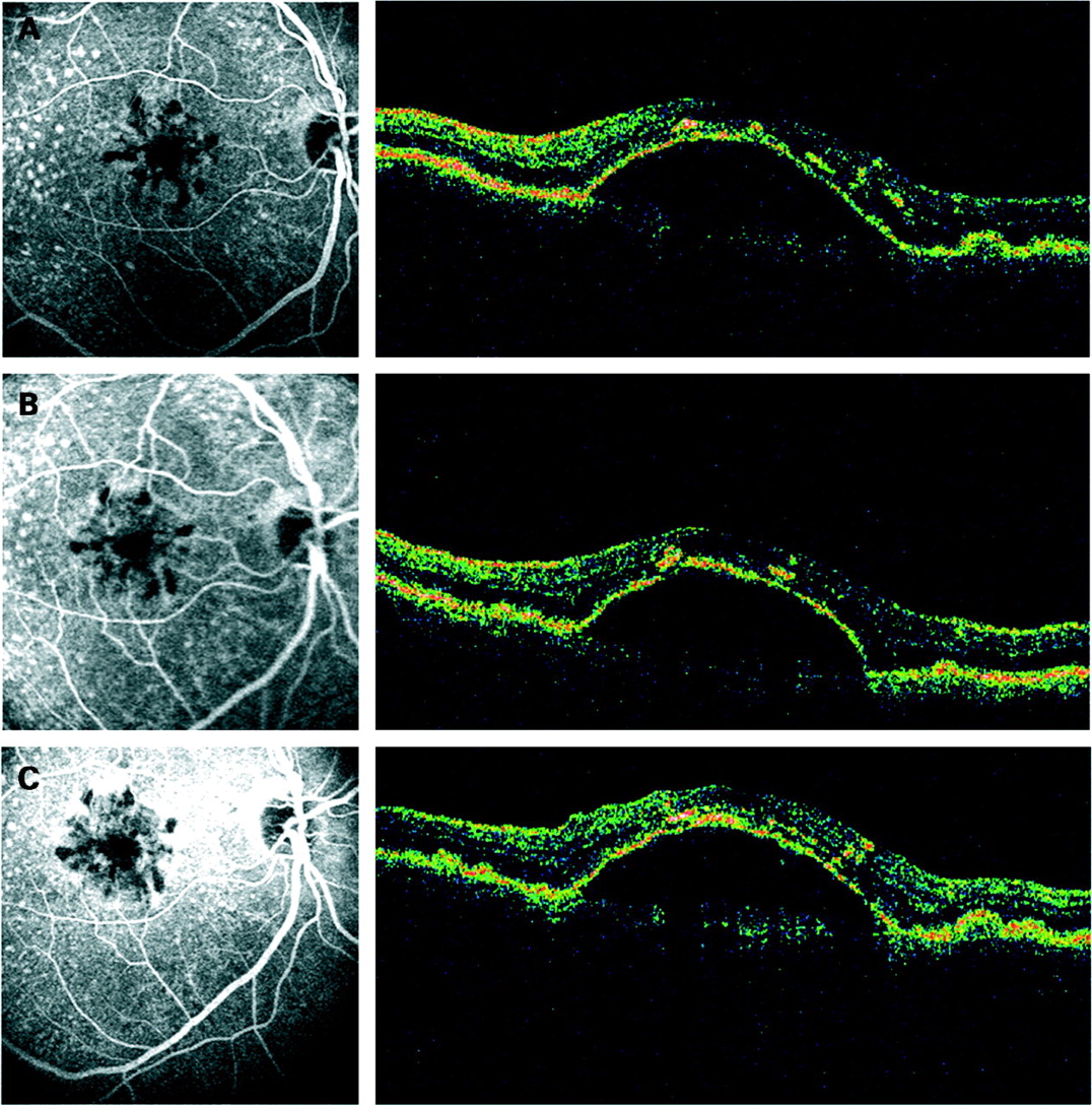
Fluorescein angiography and optical coherence tomography from the right eye of one patient who received systemic treatment with Avastin. The pictures were taken before treatment (A), after 12 weeks (B) and after 24 weeks (C). The left eye of the same patient is shown in fig 2.

Fluorescein angiography and optical coherence tomography from the left eye of one patient who received systemic treatment with Avastin. The pictures were taken before treatment (A), after 12 weeks (B) and after 24 weeks (C). The right eye of the same patient is shown in fig 1.

Fluorescein angiography and optical coherence tomography from the right eye of one patient who received NaCl. The pictures were taken before treatment (A), after 12 weeks (B) and after 24 weeks (C). The left eye of the same patient is shown in fig 4.

Fluorescein angiography and optical coherence tomography from the left eye of one patient who received NaCl. The pictures were taken before treatment (A), after 12 weeks (B) and after 24 weeks (C). The right eye of the same patient is shown in fig 3.
At the end of follow-up, after 24 weeks, the BCVA and RVA were 0.33 (0.44) and 0.3 (0.4)logRAD respectively in the Avastin group and 0.53 (0.42) and 0.5 (0.4)logRAD in the NaCl group. The macular thickness and lesion sizes were 272.3 (91.5) μm and 4.86 (1.92) mm respectively in the Avastin group (figs 1, 2), 290.8 (78.2) μm and 3.93 (1.93) mm in the NaCl group (figs 3, 4). The mean blood pressure remained 140/80 mm Hg in the NaCl group, while the systolic pressure increased to 150 (12) mm Hg, and the diastolic pressure increased to 90 (12) mm Hg in the Avastin group. The IOP was 15 (1) mm Hg in the Avastin group and 17 (2) mm Hg in the NaCl group.
After 10 weeks, the BCVA in the Avastin group dropped to 0.39 (0.46), which was a difference of 0.07 (0.06) compared with BCVA in the same group before the start of therapy. There were no differences in BCVA in the NaCl group and no differences in RVA in either group.
Throughout the 24 weeks, the macular thickness decreased in the Avastin group by 103.6 (14.9) μm, while there was an increase of 22.1 (8.4) μm in the NaCl group (table 1). In the NaCl group, the highest increase in macular thickness was found after 10 weeks, which was an increase of 133.7 (20.4) μm. In the Avastin group, the thinnest macular thickness was measured at 5 weeks, with a difference of 138 (2.6) μm.
The lesion size decreased in the Avastin group from 6.1 (2.2) mm before treatment, to 5.1 (1.9) mm at week 12 and 5.6 (2.1) mm after 24 weeks. In the NaCl group, the lesion size increased from 3.7 (1.9) mm prior to treatment, to 3.8 (2.0) mm after 12 weeks and 3.9 (1.9) at the end of follow-up.
In both the Avastin and the NaCl group, five eyes remained stable, with the BCVA being ±5 letters throughout the 24 weeks, two remained stable with ±10 letters, and one showed a decrease with −15 letters.
In the FA, all eight eyes in the NaCl group were shown to remain exudative, while in the Avastin group, four (50%) eyes became dry and fibrotic, three (37.5%) eyes remained exudative, and one (12.5%) eye had subretinal bleeding. The subretinal bleeding began in week 10, while the other eye turned fibrotic.
DISCUSSION
When Michels and coauthors published the idea of systemic Avastin treatment against AMD,6 the idea of systemic anti-VEGF treatment against AMD was implicated. The idea of treating both eyes at the same time was also very tempting, since it has been proven that patients with occult CNV with serous PED in the first eye have a significantly higher risk of visual loss in the second eye.25 A systemic treatment with an initial three infusion therapies over 6 weeks seemed very promising. Lesions too large for PDT had no treatment option, since intravitreal anti-VEGFs were not yet available, and they were thought to be very invasive bearing the risk of endophthalmitis. Subretinal surgery12–14 was too invasive for those eyes, too.
The advantage of our BEAT-AMD-Study, compared with the SANA-Study,6 is that our study is randomised, double-blinded and placebo-controlled. Neither the patient nor the investigating ophthalmologist knew which treatment arm the patient was randomised into. We began our study in August 2005 and recruited until January 2008. However, recruitment has been shown to become more difficult after the idea of the off-label use of Avastin intravitreal injections has worked itself out.17 18 Those intravitreal injections of anti-VEGFs3 5 have been shown not to be as invasive as assumed,18 only a few patients develop exudative AMD on both eyes, and of these most have some internal medical problem, which did not allow inclusion into the BEAT-AMD-Study. For those reasons, we decided to discontinue recruitment and publish these data.
Our main outcome measure was lesion size, and we were able to show a decrease of 0.5 (0.08) mm in the Avastin group throughout the 24 weeks. Macular thickness and visual acuity were additional outcome measures, whereby we found decreases in macular thickness in the Avastin group already within the first 2 weeks, with a difference of 100.7 (4.5) μm lasting until week 24. However, neither the BCVA nor the RVA showed any improvement in this group. Unfortunately, there was a decrease in BCVA in week 10 by 0.07+0.06, which may be due to the development of the subretinal bleeding in one eye of a patient in week 10. Furthermore, there was a slight rise in blood pressure in this group, which began with 140/80 mm Hg and ended with the systolic pressure being 150 (12) mm Hg and the diastolic pressure being 90 (12) mm Hg. The rise in blood pressure was controlled and treated by the specialist for internal medicine and did not exceed 160/100 mm Hg in any patient.
In the placebo control group, there was an increase in lesion size throughout the 24 weeks. BCVA and RVA remained stable. Although, after 24 weeks, macular thickness was comparable with the thickness before treatment, a large increase was found at week 10, which showed a difference of 133.7 (20.4) μm compared with prior treatment. Blood pressure remained stable.
When defining the BCVA by loss of lines, both groups had the same outcome. In both groups only one eye showed a loss of three lines (15 letters), while the other eyes remained stable. However, the FA in week 24 showed that 50% of the eyes turned dry and fibrotic, 37.5% remained unchanged and 12.5% developed a subretinal bleeding in the Avastin group, while all eight eyes remained exudative in the NaCl group. IOP remained unchanged in both groups.
Even though the macular thickness decreased in the Avastin group, the BCVA did not improve. In the NaCl group, the BCVA and the macular thickness remained stable. This might be due to the fibrovascular reaction of the macula in the Avastin group, developing a fibrotic scar, with low BCVA, while a PED with a greater macular thickness still seems to have an equivalent BCVA.
In conclusion, the lesion size and the macular thickness decreased in the Avastin group. The OCT and FA showed that the macula turns dry and fibrotic under Avastin systemic treatment. Development of BCVA and RVA is comparable in both groups and did not show any variation. Avastin has systemic side effects such as arterial hypertension and thrombosis. Due to its systemic side effects, expensive costs and comparable results with intravitreal injections we suggest offering this systemic therapy to selected patients. After careful selection, according to thrombosis anamnesis, patients with exudative AMD in both eyes, patients who refuse an intravitreal injection and patients who have had previous vitrectomy should be considered for this treatment option.
REFERENCES
Footnotes
Competing interests: None.
Funding: There was financial support by the “Scientific Grant of the Mayor of Vienna,” received in June 2006.
Ethics approval: Ethics approval was provided by the Ethics Committee of the City of Vienna.
Patient consent: Obtained.