
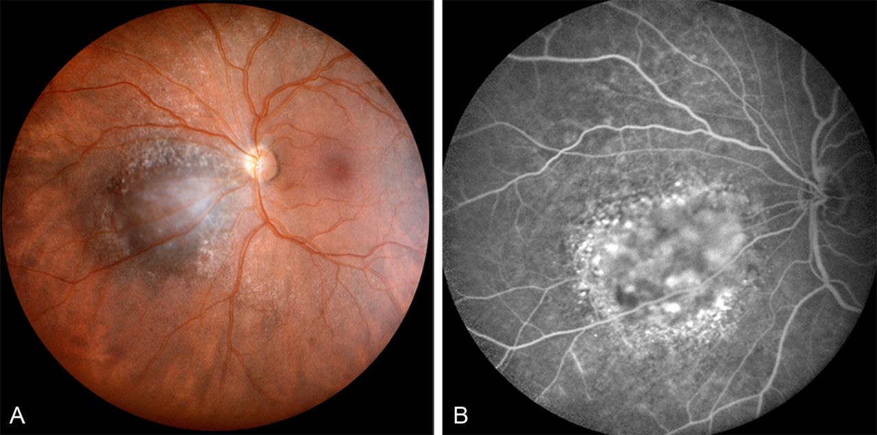
Figure 1
(A) Panoramic fundus picture (PANORET camera) of a small pigmented parapapillary choroidal tumour covered with drusen and without orange pigment at its surface. (B) On fluorescein angiography, the absence of an associated serous retinal detachment or leaking ‘pin points’ is confirmed, as well as the presence of pigment epithelial alterations at the lesion's periphery.
Vol 108 Issue 4
Table of Contents


Share this article
Click the icon of the social media platform on which you would like to share this article.


Email this article to a friend
Respond to this article