Article Text

Abstract
Aim To investigate changes in central corneal thickness (CCT) and intraocular pressure (IOP) in children after congenital cataract surgery, as well as risk factors associated with these changes.
Methods 37 eyes of 26 children with congenital cataract undergoing surgery were prospectively recruited. IOP and CCT measurements were performed before the surgery and 6, 12, 18, 24 and 36 months after the procedure.
Results Among the 37 eyes, 15 became aphakic and 22 pseudophakic. Mean CCT significantly increased from 556.24±44.19 to 585.07±56.45 μm (p=0.003) after 3 years, whereas mean IOP significantly increased from 12.05±2.3 to 13.89±2.96 mm Hg (p=0.037). Aphakic eyes underwent surgery at an early age (15.16±32.02 months) compared with pseudophakic eyes (71.48±53.14 months) (p<0.001). After 3 years, mean CCT change in aphakic eyes (56.10±46.97 μm) was significantly higher than in pseudophakic eyes (12.71±38.41 μm) (p=0.015). Age at the time of surgery was inversely correlated to CCT change (r=−0.34, p=0.04), but not to IOP change (r=−0.18, p=0.27). When surgery was performed between 0 and 1 year of age, mean CCT change at 3 years was 70.11±42.3 μm, compared with 6.27±28.09, −17.0±8.04 and 48.33±34.99 μm when surgeries were performed at 1–5, 5–10 and >10 years old, respectively (p<0.001). IOP change was not correlated to CCT change (r=0.31, p=0.06).
Conclusions CCT increases in eyes undergoing congenital cataract surgery, especially when the surgery is performed at an early age.
- Congenital cataract
- central corneal thickness
- intraocular pressure
- child health (paediatrics)
- glaucoma
- treatment surgery
- diagnostic tests/investigation
- epidemiology
- visual perception
- public health
- imaging
- lens and zonules
- contact lens
- cornea
- wound healing
- field of vision
- optic nerve
Statistics from Altmetric.com
- Congenital cataract
- central corneal thickness
- intraocular pressure
- child health (paediatrics)
- glaucoma
- treatment surgery
- diagnostic tests/investigation
- epidemiology
- visual perception
- public health
- imaging
- lens and zonules
- contact lens
- cornea
- wound healing
- field of vision
- optic nerve
Congenital cataract is an important cause of visual impairment among children. Management of congenital cataracts has changed over the last 30 years, but the occurrence of postoperative complications, such as amblyopia and secondary glaucoma, continue to threaten the visual outcome.1–4 The prevalence of glaucoma has been reported to vary from 5% to 32% depending on the follow-up time,4 ,5 reaching close to 100% in one study.6 The difficulty in establishing a precise criterion for the diagnosis of glaucoma in children,7 the use of different surgical techniques, variable follow-up and different ages at diagnosis and cataract extraction are possibly responsible for this wide range of glaucoma prevalence.5 ,8–10
Since intraocular pressure (IOP) is an important parameter for diagnosing glaucoma in children, any variable that affects IOP measurement could lead to an incorrect diagnosis of glaucoma. IOP measurements obtained with Goldmann applanation tonometer tend to be overestimated in eyes with thick corneas and underestimated in eyes with thin corneas.11 Although this information was obtained in healthy eyes without previous surgery, central corneal thickness (CCT) has been reported to be increased in aphakic and pseudophakic eyes of children compared with normal eyes.12–18 CCT abnormalities have also been reported in paediatric patients with Marfan syndrome, Down's syndrome, aniridia and osteogenesis imperfecta.19–22 Nevertheless, it is still unclear whether the increased CCT is present before cataract surgery or develops postoperatively. Most of the studies investigating this topic are retrospective,12–17 except a recent report that longitudinally analysed CCT changes after congenital cataract surgery and concluded that there is an early increase in CCT, which can be enhanced when associated with glaucoma.18
We designed a prospective study to investigate changes in CCT and IOP in children after congenital cataract surgery and to identify risk factors associated with these changes.
Methods
In all, 37 eyes of 26 children with congenital cataract scheduled to undergo surgery were prospectively recruited at the Congenital Cataract Service of the University of Campinas, Brazil, between June 2006 and January 2009. Written informed consent was obtained from all participants or a legally responsible person after approval by the Institutional Ethics Committee (number 248/2006 CAAE 0181.0.146.000-06). The study protocol followed the tenets of the Declaration of Helsinki.
Children with clinically detectable corneal oedema of any grade, history of systemic or ocular disease that could interfere with measurements (such as corneal dystrophies and sphingolipidoses), previous ocular trauma or surgery, aniridia, glaucoma and use of topical or systemic medications that could influence IOP or CCT measurements were excluded.
Demographic data such as gender, age at the time of surgery, race and ophthalmic history were collected. All subjects underwent a standard ophthalmic examination by the same observer, including best-corrected visual acuity (except when the child was too young to inform visual acuity), slit lamp biomicroscopy, applanation tonometry and dilated pupil ophthalmoscopy. Goldmann applanation tonometry was performed in individuals able to be examined at the slit lamp (model R900, Haag-Streit, Koeniz, Switzerland). Younger uncooperative children underwent IOP measurements with the Perkins tonometer (Clement Clark International, London, UK) under anaesthesia, performed with halogenated anaesthetic (sevoflurane or isoflurane) with a mask at 0.5–1.0 minimum alveolar concentration on a 50% oxygen–nitrous oxide mixture. A low dose of halogenated anaesthetic was used for a superficial level of sedation, and Perkins tonometry was performed no longer than 5 min after induction.23–25
Direct ophthalmoscopy was performed in individuals unable to cooperate with a slit lamp 78-dioptre lens examination. CCT measurements were performed in all eyes with an ultrasound pachymeter (Ocuscan RXP, Alcon Laboratories Inc., Fort Worth, Texas, USA). The mean of 10 CCT readings was used for analysis. IOP and CCT measurements were performed immediately before the surgery and 6, 12, 18, 24 and 36 months after the procedure.
The surgery was performed using phacoaspiration with curvilinear and continuous anterior capsulorrexis and IOL implantation depending on the child's age (Crystal Type 7B, Alcon Inc., Fort Worth, Texas, USA). In general, children were left aphakic before 2 years old, whereas older children received an IOL, except when there was not enough capsular support. In younger children, who were felt to be uncooperative, a posterior capsulotomy and anterior vitrectomy were performed during the surgery. Older, cooperative children underwent ND-YAG laser posterior capsulotomy when necessary. Surgeries were performed or supervised by one of two surgeons.
Glaucoma was defined as the presence of optic nerve changes indicative of glaucomatous damage associated with IOP>21 mm Hg. To be considered glaucomatous, the optic nerve had to show at least two of the following characteristics: cup to disc ratio larger than 0.6, cup to disc asymmetry >0.2, localised notch or thinning of the rim and optic disc haemorrhage.
Categorical variables were compared using the χ2 or the Fisher exact test. Continuous variables were compared using the Mann–Whitney U test or Student t test. Linear regression analyses were performed to investigate the influence of age and IOP on CCT changes. We also evaluated changes in IOP and CCT in different age groups (0–1, 1–5, 5–10 and >10 years old). p Values of <0.05 were considered statistically significant.
Results
In all, 37 eyes of 26 children were included. Mean follow-up was 30.5±10.6 months (range 6–36 months). Overall, 15 eyes (40.5%) became aphakic and 22 (59.5%) pseudophakic. One eye (2.7%) developed glaucoma after 2 years and had to undergo an Ahmed valve implantation. This child was excluded from the analysis after the diagnosis of glaucoma. One child died after 1 year of follow-up, and four children (six eyes) lost to follow-up after 1 year. Among the 22 pseudophakic eyes, 4 (18.2%) were younger than 2-years-old at surgery, whereas 2 (13.3%) of the 15 aphakic eyes were older than 2-years-old. In all, 81% of IOP assessments were made at the surgical centre in the beginning of the follow-up, while only 7% of IOP assessments were made at the surgical centre at the end of 3 years.
Among the 26 patients, 15 (57.7%) were male and 11 (42.3%) female subjects. In the aphakic group, there were significantly more male subjects (n=8, 88.9%) than in the pseudophakic group (n=7, 41.2%) (p=0.008). There was no statistically significant difference in race distribution between the phakic and pseudophakic groups (p=0.388) (table 1).
Demographics and mean follow-up in aphakic and pseudophakic groups
The mean age at the time of surgery was 48.6±53.2 months (range: from 2.1 to 178.6 months). As expected, the mean age at the time of surgery was significantly lower in the aphakic versus the pseudophakic group (15.2±32.0 and 71.5±53.1 months, respectively) (p<0.001) (table 1). There was no statistically significant difference between the mean follow-up in the aphakic and pseudophakic groups (28.4±12.3 and 31.9±9.3 months, respectively) (p=0.359).
Table 2 displays the mean CCTs for the group as a whole and for the pseudophakic and aphakic groups. Mean CCT significantly increased from 556.24±44.19 μm to 585.07±56.45 μm (p=0.003). The mean CCT change (final CCT – initial CCT) was significantly higher in the aphakic group (56.1±46.97 μm) than in the pseudophakic group (12.71±38.41 μm) (p=0.015). Among the four younger children who became pseudophakic, three showed CCT increases that varied from 12 to 105 μm at 36 months. None of the older children who became aphakic showed increased CCTs (−23 and −21 μm at 24 and 36 months, respectively).
Preoperative CCT and mean CCT change in all time intervals
Mean IOP significantly increased from 12.05±2.3 to 13.89±2.96 mm Hg (p=0.037) (table 3). There was no statistically significant difference in mean IOP change (final IOP – initial IOP) between the aphakic (2.80±4.61 mm Hg) and pseudophakic groups (1.33±4.16 mm Hg) (p=0.396).
Mean IOP change in all time intervals
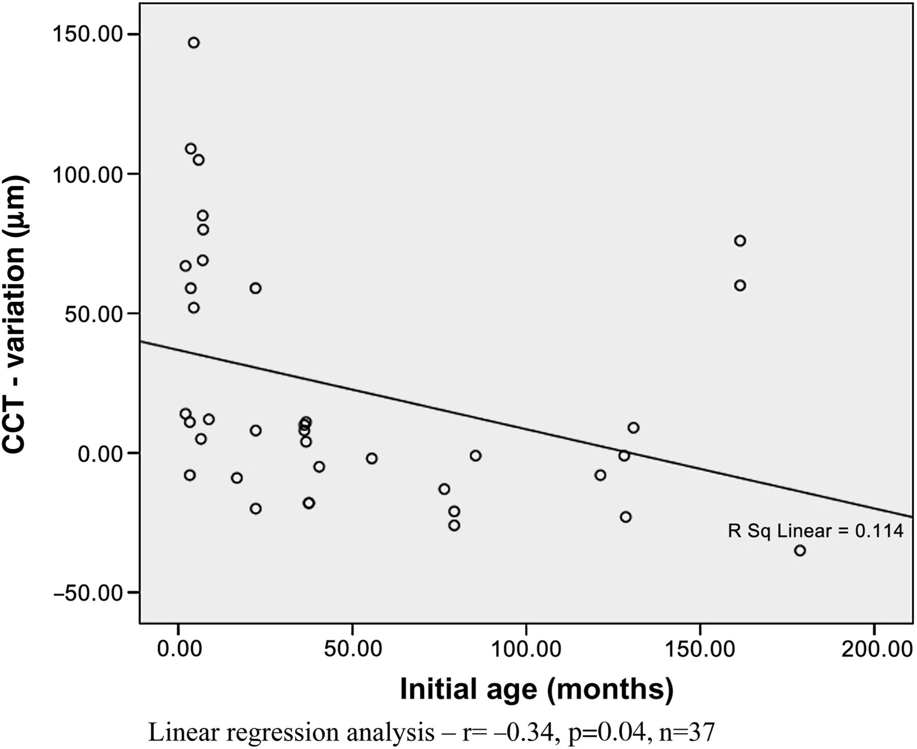
At 36 months of follow-up, mean CCT change was 70.11±42.3 μm compared with 6.27±28.09, −17.0±8.04 and 48.33±34.99 μm when surgeries were performed between 0 and 1, 1–5, 5–10 and >10 years old, respectively (p<0.001). Mean IOP change also was significantly different between these groups (4.89±2.8, 0.27±3.13, 1.80±7.01 and −1.33±0.58 mm Hg, respectively) (p=0.043). Age at the time of surgery was inversely correlated to CCT change (r=−0.34, p=0.04) (figure 1), but not to IOP change (r=−0.18, p=0.27) (figure 2).

Central corneal thickness (CCT) change × age at the moment of surgery.

Intraocular pressure (IOP) change × age at the moment of surgery.
When all patients were analysed, the correlation between CCT change and IOP change almost reached statistical significance (r=0.31, p=0.06). The CCT change was directly correlated with IOP change in the aphakic group (r=0.54, p=0.04) (figure 3), but not in the pseudophakic group (r=0.09, p=0.68) (figure 4).
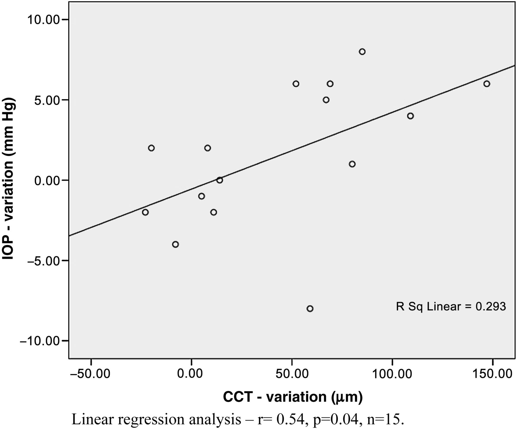
Intraocular pressure (IOP) change × central corneal thickness (CCT) change in the aphakic group.

Intraocular pressure (IOP) change × central corneal thickness (CCT) change in the pseudophakic group.
Discussion
Several retrospective studies have demonstrated that there is an increase in CCT readings in eyes following congenital cataract extraction12–18 (table 4). In some studies, aphakic and pseudophakic eyes were compared. The influence of an IOL on CCT measurements varies in the literature. Muir et al 12 observed that CCT is higher in aphakic than in pseudophakic children, a finding that was not confirmed by Simon et al.13 On the other hand, Simsek et al 14 demonstrated that secondary IOL implantation does not influence the CCT measurements. As mentioned below, this is probably explained by the different ages at the time of surgery. In some studies,13 ,14 glaucomatous and non-glaucomatous subjects were included. However, it is well known that increased IOP and the use of antiglaucomatous medications may influence CCT readings and corneal endothelial cell function.
CCT following congenital cataract extraction
In a recent short-term longitudinal study, Lim et al 18 studied 66 patients with unilateral congenital cataract and reported that CCT of both eyes were similar before cataract removal (mean CCT 552±32.9 and 550.9±40.4 μm, respectively, p=0.78). However, these measurements increased 6.5 months after cataract extraction (mean CCT increase 29.7±43.1 μm, p=0.03 in unilateral cases and 27.4±39.4 μm, p=0.01 in bilateral cases). An important limitation of this study is the fact that it included patients with glaucoma, some of them using antiglaucomatous medications. Furthermore, the short-term follow-up could not exclude a possible transient CCT increase due to corneal oedema.
Our study is one of the first longitudinal studies in the literature and has the longer mean follow-up (30.5 months) of patients undergoing congenital cataract surgery. In accordance with previously mentioned studies, we observed a CCT increase in aphakic children after cataract removal (56.1±46.97 μm) when compared with pseudophakic eyes (12.71±38.41 μm, p=0.015). Moreover, our study suggests that congenital cataract surgery performed earlier results in greater increases in CCT measurements. Two mechanisms could justify this finding: the absence of a lens could be harmful to the development of the cornea or surgeries performed at an earlier age could promote significant corneal changes.16 ,26
Our series suggests that age is probably the most important factor influencing postoperative CCT changes (figure 1). Furthermore, pseudophakic eyes operated earlier tended to present an increase of CCT measurements, whereas aphakic eyes operated later showed no increase in CCT measurements. In accordance with Lambert et al,27 younger children undergoing congenital cataract surgery tended not to receive an IOL, which explains why aphakic eyes were significantly younger than pseudophakic eyes at the time of surgery. Although the mean CCT change in the group older than 10-years-old was high (48.33±34.99 μm), the small number of patients in this group (n=3) limits the comparison with other age groups.
In normal children, the cornea thins during the early months of life and stabilises over the following years, indicating that the corneal development continues after birth during the first year of life.27 ,28 Although CCT has been shown to increase physiologically in normal children until 11 years old (varying from 553 to 573 μm in white children and Hispanic children and 541 to 551 μm in African–American children),29 the mean change observed in the aphakic group in our study is significantly greater (from 560 to 602 μm).
It is believed that the mechanical stress during cataract surgery and inflammation in the anterior chamber can damage the endothelial cells, but it is uncertain whether these injuries are sufficient to cause the corneal thickening.16 Surgical trauma to the cornea during the first months of life could impair mechanisms that regulate hydration, evaporation and transparency.30 Moreover, the developing cornea could be damaged by surgical trauma, induced by long surgical time or excessive BSS usage, corneal contact and collision of lens fragments due to turbulent flow and air bubble formation.31 ,32 It has been suggested that vitreous factors can modify the anterior segment microstructure and corneal maturation. The IOL could function as a barrier between anterior and posterior segments, avoiding corneal changes secondary to vitreous factors.26 However, our study indicates that aphakic eyes operated later do not develop corneal thickening. This fact reinforces that earlier age at surgery (and not aphakia) is the main risk factor associated with this change.33
A reduction in endothelial cell count or endothelial cell dysfunction could be responsible for corneal thickening. Nilforushan et al 15 did not observe statistically significant differences regarding the endothelial cell count characteristics between operated eyes and the control group. However, Amino et al 16 concluded that, although there was no difference in corneal endothelial cells counts among the cataract-extracted eyes and controls (p=0.36), the frequency of hexagonally shaped endothelial cells and the coefficient of variation in the endothelial size were higher in the cataract-extracted eyes (p<0.01, p<0.01). Unfortunately, our study did not include the use of specular microscopy to measure endothelial cell count.
Mean IOP was significantly increased in our study (p=0.037) but there was no correlation between IOP and age at surgery (r=−0.18, p=0.27). The CCT change was directly correlated with IOP change in the aphakic group (r=0.54, p=0.04), but not in the pseudophakic group (r=0.09, p=0.68). This fact could be explained by the higher CCT measurements obtained in aphakic children.
There are four possible explanations for the low incidence of glaucoma (2.7%) in our series. First, our mean follow-up was short compared with other studies, where the mean follow-up was higher than 5 years.4 ,10 ,33 Also, the improvement in surgical techniques (smaller incisions, decreased surgical time, posterior capsulotomy with anterior vitrectomy, use of foldable IOL at earlier ages)8 ,31 ,34 may have reduced the incidence of postoperative complications, including glaucoma. Moreover, our patients underwent surgery in a later age, and early age at surgery is a known risk factor for the development of glaucoma in eyes undergoing congenital cataract surgery. Finally, we excluded eyes with persistent fetal vasculature, a risk factor for glaucoma, from our series.35 Furthermore, the diagnostic criteria included optic nerve alterations, which was not the case for most of the previous studies that relied only in IOP. However, it is possible that subjects with elevated IOP and no optic nerve damage that were included as non-glaucomatous in our series may actually develop optic nerve changes in the future.
Although some individuals had IOP measurements performed with Perkins tonometry and others with Goldmann applanation tonometry, previous studies had shown no significant difference between their measurements.36 ,37 It has been shown that anaesthesia may reduce IOP in children,25 but we used a low concentration of halogenated anaesthetic and performed IOP measurements within 5 min after induction.
Functional and anatomical evaluations may be difficult to perform in children with congenital cataract or aphakia due to nystagmus, amblyopia and cooperation. Hence, IOP measurement is an important diagnostic variable for glaucoma.5–7 In this study, we have demonstrated that eyes undergoing congenital cataract surgery at an early age show thicker corneas after the procedure. These CCT increases (up to 147 μm) could be clinically significant and may artificially increase IOP measurements obtained with applanation tonometry.38
References
Footnotes
-
Meeting Presentation Presented in part at the World Glaucoma Congress 2011 (Paris, France) and Pan-American Congress of Ophthalmology (Buenos Aires, Argentina).
-
Competing interests None.
-
Patient consent Obtained from the parents.
-
Ethics approval Ethics approval was provided by the Institutional Ethics Committee (number 248/2006 CAAE 0181.0.146.000-06).
-
Provenance and peer review Not commissioned; externally peer reviewed.
-
Correction notice This article has been corrected since it was published Online First. The author name 'Vital Costa' has been updated to read 'Vital P Costa'.
Linked Articles
- At a glance
- PostScript