Article Text

Abstract
AIMS Slit lamp fundus biomicroscopy allows for high magnification, stereoscopic diagnosis, and treatment of macular diseases. Variable contrast, narrow field of view, and specular reflections arising from the cornea, sclera, and examining lens reduce image quality; these images are of limited clinical utility for diagnosis, treatment planning, and photodocumentation when compared with fundus camera images. Algorithms are being developed to segment fundus imagery from slit lamp biomicroscopic video image sequences in order to improve clinical utility.
METHODS Video fundus image sequences of human volunteers were acquired with a video equipped, Nikon NS-1V slit lamp biomicroscope. Custom developed software identified specular reflections based on brightness and colour content, and extracted the illuminated fundus image based on colour image analysis and size constraints.
RESULTS In five subjects with variable image quality, the approach allowed for automatic, robust, accurate extraction of that portion of the video image corresponding to the illuminated portion of the fundus. Non-real time analysis allowed for fundus image segmentation for each frame of the image sequence. In real time, segmentation occurs at 2 Hz, and improvements are being implemented for video rate performance.
CONCLUSIONS Computer vision algorithms allow for real time extraction of fundus imagery from marginal quality, slit lamp fundus biomicroscope image sequences.
- computer vision algorithms
- slit lamp
- fundus images
Statistics from Altmetric.com
Slit lamp fundus biomicroscopy allows for high magnification, stereoscopic diagnosis, and treatment of macular diseases. However, variable contrast, narrow field of view, and specular reflections arising from the cornea, sclera, and examining lens reduce image quality.
While there has been great interest and much effort directed towards analysis, feature extraction, change detection, and image guided treatment based on high quality fundus images,1-9computerised analysis of slit lamp fundus biomicroscopic images has received little attention, perhaps in part attributable to the presumed difficulties in extracting signal (fundus features) from noise (other image components and reflections). As the workhorse for ophthalmic diagnosis and treatment, the slit lamp biomicroscope is ubiquitous. Alternately, fundus cameras are more expensive and less readily available. Therefore, our group and others have been exploring the application of image processing methodologies towards increasing the clinical utility of acquired slit lamp biomicroscopic images for diagnosis, treatment planning, and photodocumentation.10-12
In this report, we develop and evaluate algorithms as a first step toward allowing for enhancement, processing, and analysis of slit lamp fundus biomicroscopic images.
Methods
IMAGE ACQUISITION
Subjects were derived from the clinical practice of the retina service at the Scheie Eye Institute. With appropriate institutional review board approval, and following informed consent, slit lamp biomicroscopic fundus examinations were performed with a video equipped, Nikon NS-1V slit lamp biomicroscope using a Goldmann three mirror contact lens or a 78 dioptre non-contact, hand held lens. A beam splitter attached to one of the oculars permitted image capture by a charge coupled device (CCD) camera. Live image analysis could be performed. For development purposes and subsequent analyses, the examinations were stored at 30 frames per second on S-VHS videotape.
IMAGE PROCESSING
The video signal was sent to a Pentium III, 600 MHz personal computer with frame grabber and digitiser (Matrox Corona, Quebec, CA, USA). Custom developed software identified specular reflections based on brightness and colour content, and extracted the illuminated fundus image based on colour image analysis and size constraints.
While the fundus image has little blue colour content, specular reflections from the cornea, sclera, and contact lens were noted to have significant brightness contributions in the green and blue channels. The illuminated portion of the fundus in the slit lamp biomicroscope image is noted to correspond to the single, contiguous, vertically elongated blob of pixels with brightness in green and red channels, but with a small blue channel component.
Results
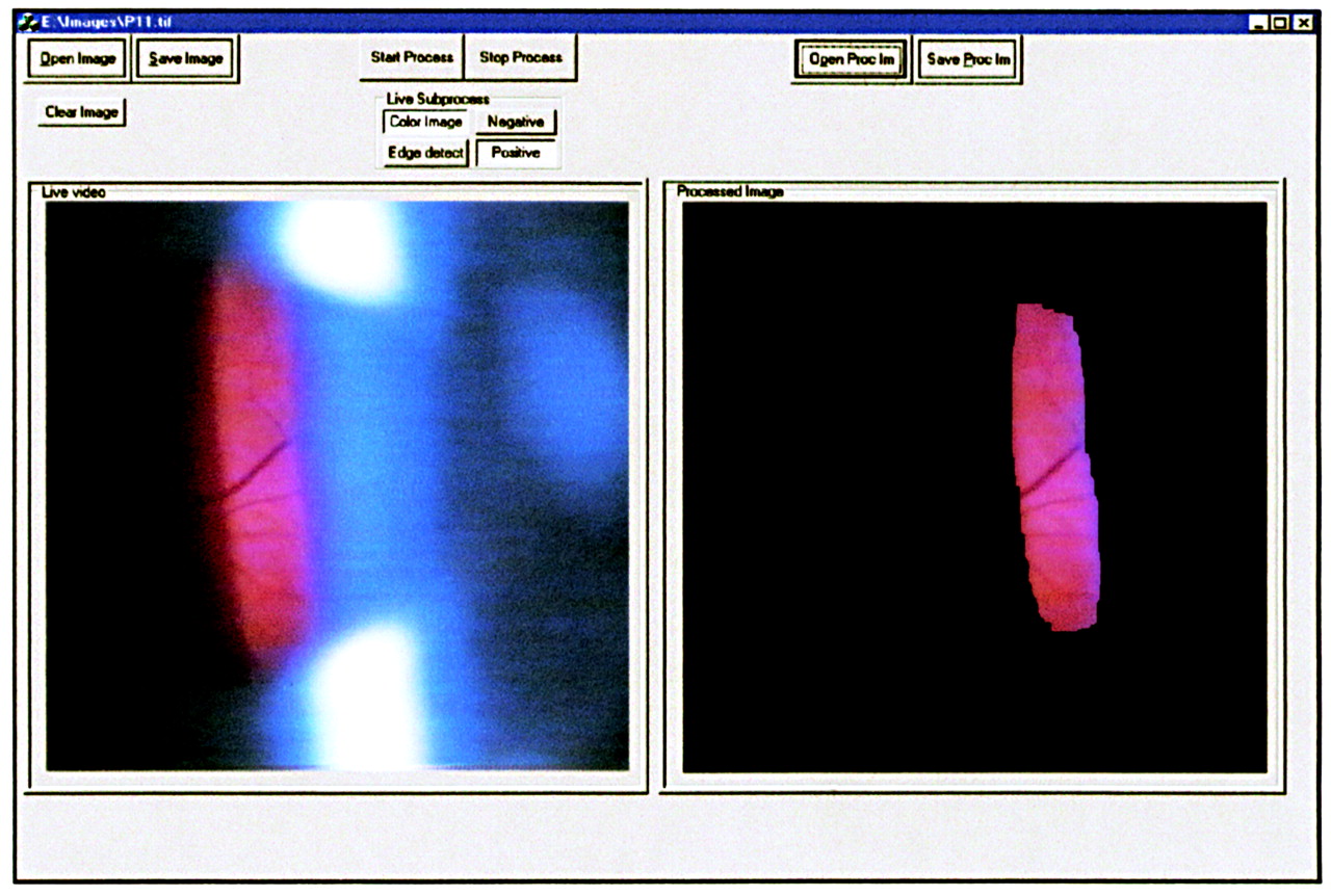
On evaluation in five subjects with variable image quality, our approach allows for robust identification of specular reflections and automatic extraction of that portion of the video image corresponding to the illuminated portion of the fundus. Figure 1 (left) depicts a slit lamp fundus biomicroscopic image with usual specular reflections. Figure 1 (right) depicts the processed image, corresponding to the illuminated portion of the fundus. Stray reflections have been eliminated. The process is run in a graphical user interface (GUI) environment; generated images and image sequences are depicted side by side with the incoming video stream for comparison. Images and image sequences can be stored for subsequent analysis, review, and processing.

Graphical user interface environment for real time extraction of fundus images. (Left) Unprocessed, digitised slit lamp biomicroscopic fundus image. The fundus image subtends a small fraction of the total image area, and specular reflections degrade the overall image quality. (Right) Automated real time fundus image extracted from the unprocessed image. The image content corresponding to the fundus image is preserved, while extraneous content and specular reflections are eliminated.
Non-real time analysis allows for fundus image segmentation for each frame of the image sequence. In real time, segmentation occurs at 2 Hz, and improvements are being implemented for video rate (30 Hz) performance. Successful segmentation was possible with poor image quality and suboptimal focus.
Discussion
While slit lamp fundus biomicroscopy is the usual method for high magnification inspection of fundus details, variable image quality, specular reflections, and narrow field of view reduce the utility of this approach for diagnosis, treatment planning, and photodocumentation.
In a separate thrust, we have developed methods for automated and semiautomated registration of slit lamp fundus images in order to improve image quality through oversampling and signal averaging, as well as expand the otherwise limited field of view through image montaging (Berger et al, unpublished). In addition, extraction of useful imagery will allow for revealing biomicroscopic photographic correlation as is necessary for image comparison, biomicroscopic angiographic correlation as is required for laser treatment planning, and eventually for an image overlay environment where the slit lamp biomicroscope achieves pluripotential function for image acquisition, image comparison and correlation, as well as treatment planning and execution.10-12
The developed computer vision algorithms allow for real time extraction of fundus imagery from marginal quality, slit lamp fundus biomicroscope image sequences, and this step will facilitate further progress towards increasing the clinical usefulness for this platform for image acquisition and analysis.
Acknowledgments
Supported by a career development award, Research to Prevent Blindness (JWB) and K-08 00374 (JWB).