Article Text

Abstract
BACKGROUND Pulsatile ocular blood flow (POBF) is a parameter for evaluating choroidal blood flow. POBF in the patients with non-exudative and exudative age related macular degeneration (AMD) was investigated.
METHODS POBF, pulse amplitude (PA), systolic and diastolic blood pressures, intraocular pressure (IOP), refractive error, and axial length were compared among 10 patients with non-exudative AMD, 11 patients with exudative AMD, and 69 age matched controls. A Langham OBF computerised tonometer was used with the participants in the sitting position to measure POBF and PA.
RESULTS No significant differences were found in age, systolic and diastolic blood pressures, IOP, or refractive error between patients with exudative and non-exudative AMD and the control subjects. In the patients with exudative AMD the POBF (median, 372.7 μl/min) and PA (median, 1.2 mm Hg) were significantly lower than in the patients with non-exudative AMD (median, 607.0 μl/min (p = 0.02) and 2.2 mm Hg (p = 0.04), respectively) and control subjects (median, 547.4 μl/min (p = 0.01) and 2.0 mm Hg (p = 0.01), respectively).
CONCLUSIONS These data show that the POBF and PA in the patients with exudative AMD are lower than in the patients with non-exudative AMD and normal subjects. Decreased choroidal blood flow may have a role in the development of choroidal neovascularisation in AMD.
- age related macular degeneration
- choroidal blood flow
- pulsatile ocular blood flow
- pulse amplitude
- choroidal neovascularisation
Statistics from Altmetric.com
- age related macular degeneration
- choroidal blood flow
- pulsatile ocular blood flow
- pulse amplitude
- choroidal neovascularisation
Age related macular degeneration (AMD) is the major cause of irreversible blindness in elderly patients worldwide.1-4The pathogenesis of this disease has been investigated from genetic,5 histological,6 and haemodynamic perspectives,7-10 but it is poorly understood. AMD is classified as non-exudative, the dry type, or exudative neovascular, the wet type.1112 Choroidal neovascularisation (CNV) in the macula causes severe visual impairment in AMD. Vascular endothelial growth factor (VEGF) and other angiogenetic factors, which are induced by hypoxia and ischaemia, may play a part in the development of CNV.13-15 Choroidal circulation may be important for the development of CNV, the late stage of AMD.
Several authors have evaluated ocular blood flow in patients with AMD.7-10 However, the choroidal blood flow is uncertain in patients with non-exudative and exudative AMD. Determining pulse amplitude (PA) and pulsatile ocular blood flow (POBF) by measuring intraocular pressure (IOP) using the Langham OBF computerised tonometer is a way to evaluate choroidal blood flow.16-18 In the current study, we evaluated the total choroidal blood flow in the patients with non-exudative and exudative AMD by measuring POBF.
Materials and methods
Patients and control subjects from the department of ophthalmology of Asahikawa Medical College were enrolled in this study. Only one eye of each participant was included. The eyes were divided as follows into three groups based on the findings of slit lamp biomicroscopy, indirect ophthalmoscopy, fundus photography, and fluorescein angiography: 69 age matched controls, 10 patients with non-exudative AMD, and 11 patients with exudative AMD. The exudative and non-exudative forms of AMD have been defined previously.1112 Subjects with any other ocular disease were excluded. Subjects with systemic hypertension were included in this study.
Informed consent was obtained in all cases. The study protocol was reviewed by the ethics committee of our institution. All procedures adhered to the tenets of the Declaration of Helsinki.
POBF and PA were measured by the Langham OBF computerised tonometer (Langham Ophthalmic Technologies, Timonium) with patients in the sitting position. Data were collected for POBF and PA for each subject, and the mean measurements were calculated from five representative pulses. Systolic and diastolic brachial arterial blood pressures were measured with a sphygmomanometer. IOP was measured with a non-contact tonometer (CT-90, Topcon, Japan). The refractive error was expressed as a spherical equivalent refraction obtained by autokeratorefractometer (KR-7100, Topcon). Axial length was measured using an A-scan biometric ultrasound, OcuScan (Alcon, Fort Worth).
POBF, PA, age, systolic and diastolic blood pressures, IOP, refractive error, and axial length were compared in the three groups. All comparisons were performed using the Mann-Whitney test. A p value <0.05 was considered statistically significant.
Results
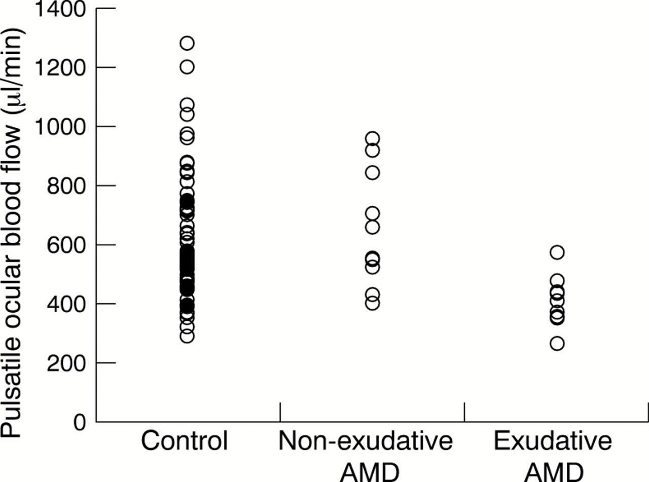
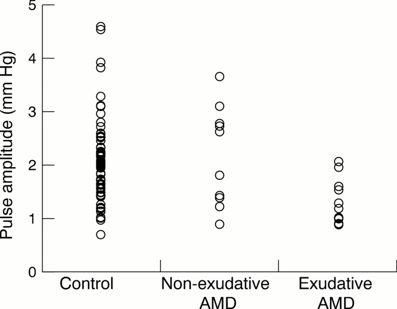
No significant differences were found in age, systolic and diastolic blood pressures, IOP, refractive error, and axial length between patients with AMD and the control subjects (Table 1). In the patients with exudative AMD, the PA (median 1.2 mm Hg; range 0.9–2.1) was significantly lower than in patients with non-exudative AMD (median 2.2 mm Hg; range 0.9–3.7; p = 0.04) and age matched controls (median 2.0 mm Hg; range 0.7–4.6; p = 0.01) (Fig 1). In patients with exudative AMD, the POBF (median 372.7 μl/min; range 261.6–570.2) was significantly lower than in patients with non-exudative AMD (median 607.0 μl/min; range 401.0–959.0; p = 0.02) and age matched controls (median 547.4 μl/min; range 290.7–1282.9; p = 0.01) (Fig 2). No significant differences were found in PA and POBF between patients with non-exudative AMD and the age matched controls (p = 0.96 and p = 0.69, respectively) (Figs 1 and2).
Characteristics of controls and patients with age related macular degeneration (AMD)

Pulse amplitude in patients with exudative age related macular degeneration (AMD) (n=11), patients with non-exudative AMD (n=10), and age matched controls (n=69).
Pulsatile ocular blood flow in patients with exudative age related macular degeneration (AMD) (n=11), patients with non-exudative AMD (n=10), and age matched controls (n=69).
Discussion
This study demonstrates that the PA and POBF in patients with exudative AMD are significantly lower than in controls. Several authors reported that the blood velocities of the short posterior ciliary artery are lower in the patients with AMD.89 These results indicate that the total choroidal blood flow is lower in the patients with exudative AMD than in controls. A prolonged choroidal filling phase on fluorescein angiography is related to thickening of the Bruch's membrane.19 The thickening of the Bruch's membrane may induce increased vascular resistance of the choroid and decreased choroidal blood flow in the patients with AMD.
Grunwald et al reported that the choroidal blood flow in the centre of the fovea was lower in the patients with non-exudative AMD than in age matched controls, primarily because of decreased blood volume measured by laser Doppler flowmetry.7 However, our results showed that POBF did not decrease in the patients with non-exudative AMD compared with age matched controls. Thus, our results suggest that only local choroidal blood flow in the centre of the fovea may decrease, but total choroidal blood flow does not decrease in the patients with non-exudative AMD compared with age matched controls.
Friedman et al reported that scleral rigidity increases and the choroidal blood flow decreases in the patients with AMD.9-21 Those authors concluded that the eyes of patients with AMD had decreased choroidal blood flow and increased resistance in the choroidal vasculature. Several investigators reported that scleral rigidity affects POBF.2223 We believe that we need to consider the relation between scleral rigidity and POBF in the patients with AMD in the future.
Lang et al showed that the POBF decreased in the patients with non-exudative AMD compared with controls.10 In our data, axial length was a major factor affecting POBF in normal subjects.24 When evaluating the choroidal blood flow by measuring POBF, the effect of axial length should be considered. In the present study, no significant differences were found in the axial length in any group. We concluded that the total choroidal blood flow in patients with exudative AMD was lower than the patients with non-exudative AMD and age matched controls.
CNV in the macula causes severe visual impairment in the late stage of AMD. Basic fibroblast growth factor (bFGF) has been detected in CNV membranes in the patients with AMD.15 In an experimental model of CNV, investigators observed a defect in Bruch's membrane and increased expression of VEGF in the accumulating macrophages and migrating retinal pigment epithelial cells and Müller cells.14 In the vitreous of patients with CNV, the levels of VEGF increased.25 Adamis et al indicated that retinal pigment epithelial cells can promote endothelial proliferation via the production and release of VEGF during hypoxia.26 These reports suggest that angiogenic factors may play a part in the formation of CNV. In the present study, POBF in patients with AMD decreased by about 30% compared with controls and patients with non-exudative AMD. The decreased total choroidal blood flow may induce CNV via angiogenic factor induced by hypoxia in the patients with exudative AMD. Further studies of choroidal circulation, which may be important for the development of CNV in the late stage of AMD, are needed.
Acknowledgments
The authors have no proprietary interest in any aspect of this technology.