Article Text

Abstract
Purpose: Carbonic anhydrase enzymes (CAs) are universally involved in many fundamental physiological processes, including acid base regulation and fluid formation and movement. In glaucoma patients, CA inhibitors are very effective in lowering intraocular pressure by reducing the rate of aqueous humour secretion mediated by the CAs in the ciliary epithelium. In this work, we investigated the expression and tissue distribution of two recently discovered CA genes CA9 (CAIX) and CA12 (CAXII) in fetal, neonatal, and adult human eyes with and without glaucoma.
Methods: CAIX and CAXII expression in 16 normal and 10 glaucomatous eyes, and in cultured non-pigmented ciliary epithelial cells (NPE) from normal and glaucoma eye donors was assessed by immunostaining. In addition, northern blot hybridisation was performed to assess expression of CA4, CA9, and CA12 mRNA in cultured NPE cells from normal and glaucoma donors.
Results: CAXII was localised primarily to the NPE with its expression prominent during embryonic eye development but which decreased significantly in adults. CAIX expression in the NPE was very low. The epithelium of cornea and lens occasionally expressed both enzymes at low levels during development and in adult eye, and no expression was detected in the retina. The NPE from glaucoma eyes expressed higher levels of CAXII, but not CAIX, in comparison with normal eyes. This expression pattern was retained in cultured NPE cell lines. NPE cells from a glaucoma patient showed a five-fold increase in the CA12 mRNA level with no detectable expression of CA9 mRNA. Also, no expression of the CA4 gene encoding a GPI anchored plasma membrane protein was detected on these northern blots.
Conclusions: Transmembrane CAIX and CAXII enzymes are expressed in the ciliary cells and, thus, may be involved in aqueous humour production. CA12 may be a targeted gene in glaucoma.
- carbonic anhydrases
- eye humour
- glaucoma
- CA, carbonic anhydrase (Arabic numerals denote the gene and corresponding mRNA while the Latin numerals denote the protein products)
- NPE, non-pigmented epithelial (ciliary) cells
- GPI, glycosylphosphatidylinositol lipid anchor
- ACG, angle closure glaucoma
Statistics from Altmetric.com
- CA, carbonic anhydrase (Arabic numerals denote the gene and corresponding mRNA while the Latin numerals denote the protein products)
- NPE, non-pigmented epithelial (ciliary) cells
- GPI, glycosylphosphatidylinositol lipid anchor
- ACG, angle closure glaucoma
Carbonic anhydrase enzymes (CAs) are universally involved in many fundamental physiological processes, including acid base regulation and fluid formation and movement.1 The involvement of carbonic anhydrases (CA) in aqueous humour secretion by the ocular ciliary epithelium was suggested by several types of experiment2,3 and directly supported by clinical observations of the efficacy in reducing raised intraocular pressure (IOP) in glaucoma by inhibitors (topical and systemic) of CA (CAinh).4–7 The clinical efficacy of CAinh plus some experimental data with isolated ciliary epithelial bilayers in vitro suggested both cytosolic and membraneous CAs mediate these effects to reduce aqueous inflow in the eye.2,7–9 Previously, cytosolic (CAII) and membrane GPI anchored (CAIV) enzymes were discovered in different anatomical and cellular sites of the eye.1,2,7 It was suggested they play a fundamental role in shifting protons and bicarbonate and consequently other solutes across membranes, thereby regulating acid base homeostasis and fluid movements.8 However, the CAIV enzyme was not found in the ciliary epithelium,3 suggesting that other membrane associated CAs might be involved in aqueous humour secretion and movements.
Recently, we and others discovered a novel class of cell surface transmembrane carbonic anhydrase genes, CA910,11 and CA12,12,13 and showed their expression in a variety of specialised adult normal human tissues and ubiquitous over-expression/induction in human cancers.12–14 In a follow up study, we examined the expression and distribution of these proteins by immunostaining tissue sections from human eye donors. In addition, we performed a northern blot analysis of cultured ciliary NPE cells15 in order to measure CA4, CA9, and CA12 mRNA levels in these cell populations.
Here we show, by immunostaining with specific antibodies, the expression of CAIX and CAXII cell surface transmembrane proteins in different anatomical structures of the human eye, namely, in embryonic, neonatal/infant, and adult eyes, under normal and pathological conditions including angle closure glaucoma. In addition, by northern blot analysis we show the expression of these genes in cultured human ciliary non-pigmented epithelial (NPE) cells from normal and glaucomatous eyes.
MATERIAL AND METHODS
Tissue specimens and cultured cells
A total of 26 eyes were collected from the pathology department at UCI Medical Center (Irvine, CA), St Joseph Hospital (Orange, CA), and San Diego Eye Bank (San Diego, CA). Among 26 eyes studied, 16 were normal eyes with no clinical or histological evidence of glaucoma and 10 were glaucomatous. Thirteen of the 16 normal eyes were from donors, and the remainder were enucleated because of extraorbital tumour (n=2) and trauma (n=1). The 10 glaucomatous eyes were enucleated because of intraorbital tumour/inflammation (n=3) and clinically diagnosed glaucoma with uncontrolled eye pain and/or blindness (n=7). The donor’s eyes were obtained either immediately after brain death or within 48 hours after death. The age distribution of the glaucoma cases ranged from 57 to 85 years, with one female of 39 years. All of the cases were diagnosed with ACG (angle closure glaucoma). As far as we could ascertain there were no hereditary cases. The donors included fetal eyes (n=5) with a gestational age of 15 to 20 weeks, neonatal/infant eyes (n=5) with an age of 1 day to 18 months, and adult eyes (n=3). All enucleated eyes were fixed in 10% neutral buffer formalin, paraffin embedded, sectioned, and stained with haematoxylin and eosin (H&E) for light microscopic examination. The study was performed with the approval of the ethics committees of each institution involved in this project and, as far as it applies, followed the tenets of the Declaration of Helsinki. Establishment and culturing of human non-pigmented ciliary epithelial cell lines (NPE) from normal and glaucoma eye donors have been described in detail previously.15 For immunostaining, NPE cell subcultures were grown in chamber slides and then fixed in a solution with one part of acetone and one part of methanol.
Immunohistochemical studies
The mouse monoclonal antibody (MN75) used to detect the MN/CAIX protein and the rabbit polyclonal antibody to CAXII protein have been described previously.14 Immunohistochemical staining of tissue sections and acetone/methanol fixed cultured cells with anti-CAIX and anti-CAXII antibodies was done using a peroxidase technique as described previously.14 Microwave pretreatment was applied to all tissue sections. Known positive and negative tissue specimens were included in each run.14 For CAIX immunostaining, the primary antibody was used at a 1:10 000 dilution and the CAXII at a 1:300 dilution. Positive staining was scored when there was plasma membrane and/or cytoplasmic brown colour reaction, and a negative score was given to tissue sections and culture cells that had no evidence of specific immunostaining.
RNA ANALYSIS
For mRNA isolation NPE cells were grown in DMEM+10% fetal calf serum to confluence and used before passage 13. mRNA isolation from cultured cells, RNA electrophoresis, and northern analysis were done as described previously.14 Quantification of northern hybridisation signals was performed using Cyclone Storage Phosphor System and OptiQuant Image Analysis Software (Packard, Meriden, CT). DNA probes representing CA9 and CA12 ORFs were obtained as described previously.14 The CA4 ORF probe was isolated from EST clone IMAGE: 2917959 (Research Genetics, Huntsville, AL) after verification of the insert by sequencing. Northern blots containing polyA+ mRNA of ciliary NPE cells from a normal two year old subject (ODM-C4, passage 9), a glaucoma patient (GCE-T, passage 12), and for comparison from the renal carcinoma cell line, 786-0,12 were hybridised sequentially with the ORF probes indicated. 28S rRNA levels were used to ensure equivalent loading of mRNA in all lanes.
RESULTS AND DISCUSSION
To identify the CAs expressed in the ciliary epithelium, we first analysed the expression of the CAIX and CAXII enzymes by immunostaining tissue sections of 26 normal and glaucomatous eyes. Microscopically, all eyes from donors had normal histology. Three non-glaucomatous eyes enucleated for extraorbital tumour/trauma also contained well preserved ciliary bodies with open angles and relatively unremarkable cornea, lens, choroid plexus, retina, sclera, and optic nerve. All glaucomatous eyes (n=10) had closed angles. Among these, two were associated with an orbital tumour and one was the result of inflammation. The rest were from patients with a clinical diagnosis of angle closure glaucoma (ACG) with no known associated disease. Histological sections of the glaucomatous eyes showed the formation of peripheral anterior synechiae, fibrosis, and degeneration of meshwork. Optic atrophy and variable degrees of degeneration of cornea and retina were observed in all glaucomatous eyes.
In normal developing eyes (gestational age ranging from 15 weeks to 20 weeks), the non-pigmented ciliary epithelium, the corneal epithelium and endothelium, and the lens epithelium expressed both CAIX and CAXII (table 1). While expression of CAXII was prominent, the expression of CAIX in the ciliary epithelium was very weak and the positive staining was limited to a few epithelial cells (fig 1A, B). In contrast, very low levels of CAXII, but no CAIX immunoreactivity, were observed along the inner membrane (the terminations of the processes of Muller’s cells) of the retina near the ora serrata. After birth the inner membrane of the retina no longer expressed CAXII and the intensity of CAXII immunoreactivity in the epithelium of the cornea, lens, and ciliary body was decreased. In adult eyes there was a persistent expression of CAXII in the non-pigmented epithelium of the ciliary body but the levels of expression were significantly decreased when the intensity of staining was compared with the developing eyes. In addition, the positive immunostaining was focal. In contrast, there was no CAIX immunoreactivity detected in the ciliary epithelial cells in the adult globes (fig 1C, D). The epithelium of the cornea and lens occasionally expressed CAIX and CAXII but the intensity of positive immunostaining was extremely weak. There was no CAIX/CAXII immunostaining in the retina.
CAIX and CAXII expression in developing, adult, and glaucoma eyes
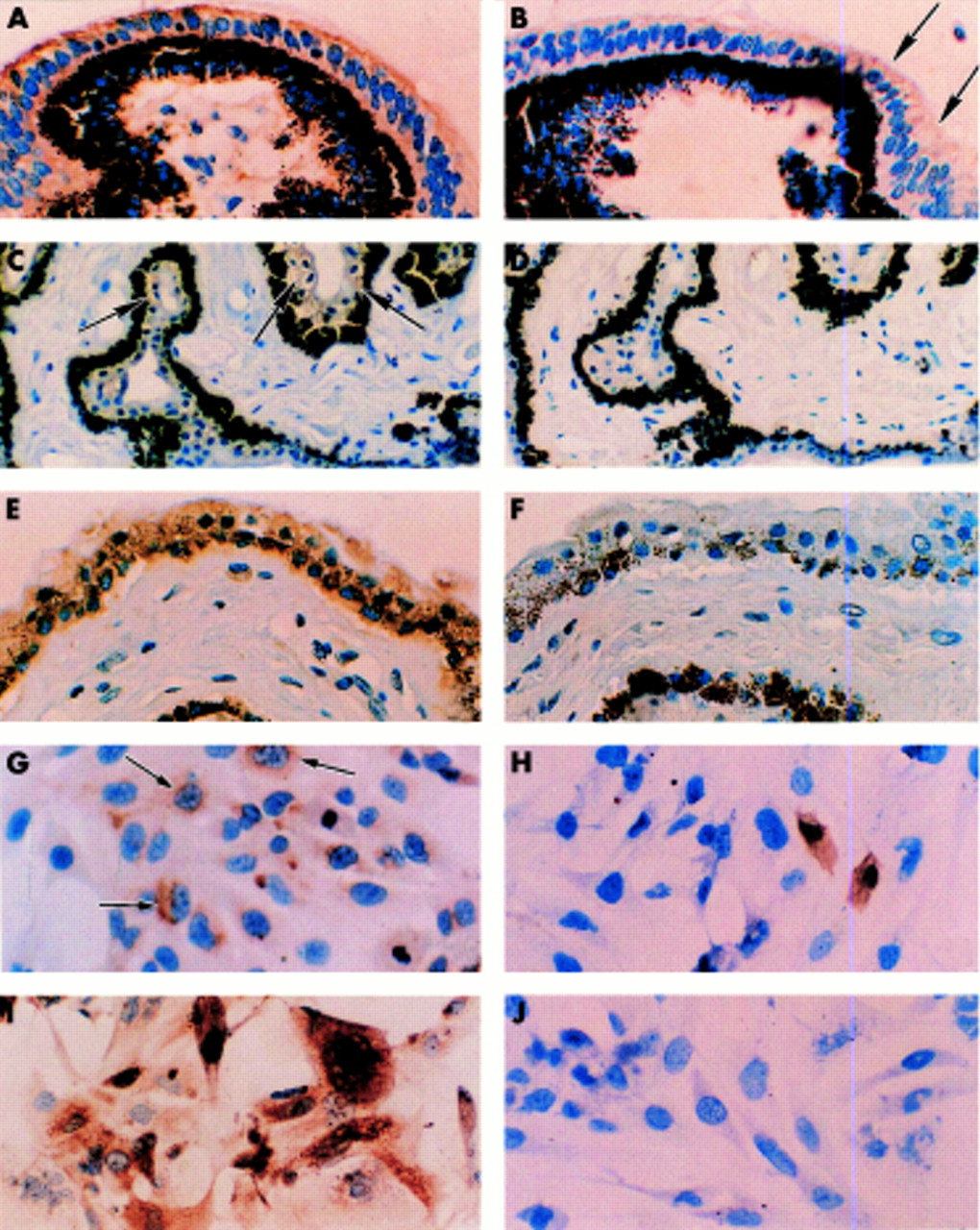
Examples of immunostaining of CAIX and CAXII proteins in non-pigmented epithelium of the ciliary body of developing (19 weeks’ gestational age), adult, and glaucomatous eyes (A-F) and of ciliary non-pigmented epithelial cultured cells of normal and glaucomatous eyes (G-J). CAXII is illustrated in A, C, E, G, and I. Immunostaining for CAIX is illustrated in B, D, F, H, and J. Diffuse immunoreactivity for CAXII and focal, very weak positivity for CAIX is seen in the developing eyes (A, B, arrows). In the fully developed eyes (adult) CAXII positive staining is weak and limited to a few cells; no CAIX immunoreactivity is seen (C, arrows, D). In contrast, high levels of CAXII expression is seen particularly in the non-pigmented ciliary epithelial cells of the glaucomatous eyes with no expression of CAIX (E, F). CAIX/CAXII expression is also seen in the cultured normal NPE cells (G, arrows, H). In the cultured glaucoma ciliary cells the intensity of CAXII immunostaining is much stronger with staining seen both in the cytoplasm and the plasma membrane. Conversely, no expression of CAIX was seen (I, J).
In glaucomatous eyes, variable degrees of CAIX/CAXII expression were observed in the epithelium of cornea and lens but the positive immunoreactivity was weak and focal. The most striking finding was high levels of CAXII, but no CAIX expression in the non-pigmented ciliary epithelium (fig 1E, F). The positive CAXII immunostaining was diffuse and the intensity of staining was moderate to strong. Clearly, CAXII expression was preferentially seen in the NPE cells. However, the high pigmentation observed in the pigmented layer precluded precise quantification of CAXII expression in the NPE cells of the ciliary bilayer. Another interesting observation was the expression of CAIX but not CAXII in the proliferative neuroglial cells of the retina in which there was severe loss of inner and outer nuclear layers. The positive staining was either focal or diffuse. A summary of the distribution of the expression of CAIX/CAXII in developing eyes before and after birth, adult donor/non-glaucomatous, and glaucomatous eyes is given in table 1.
We next determined whether these patterns of expression would be preserved in cultured NPE cells.15 The cell cultures were grown in the chamber slides and immunostained. In the normal cultured NPE cells, limited numbers of cells showed weak CAXII immunoreactivity. Even fewer cells expressed CAIX, although the level of immunostaining was somewhat stronger than CAXII (fig 1G, H). In contrast, the ciliary NPE cells from the glaucomatous eye showed high levels of CAXII expression consistent with the data obtained with the immunostained eye sections. There was a significant increase in the numbers of cell stained and in the intensity of immunostaining. However, CAIX immunoreactivity was no longer detected in the NPE cells derived from the glaucomatous eye (fig 1I, J).
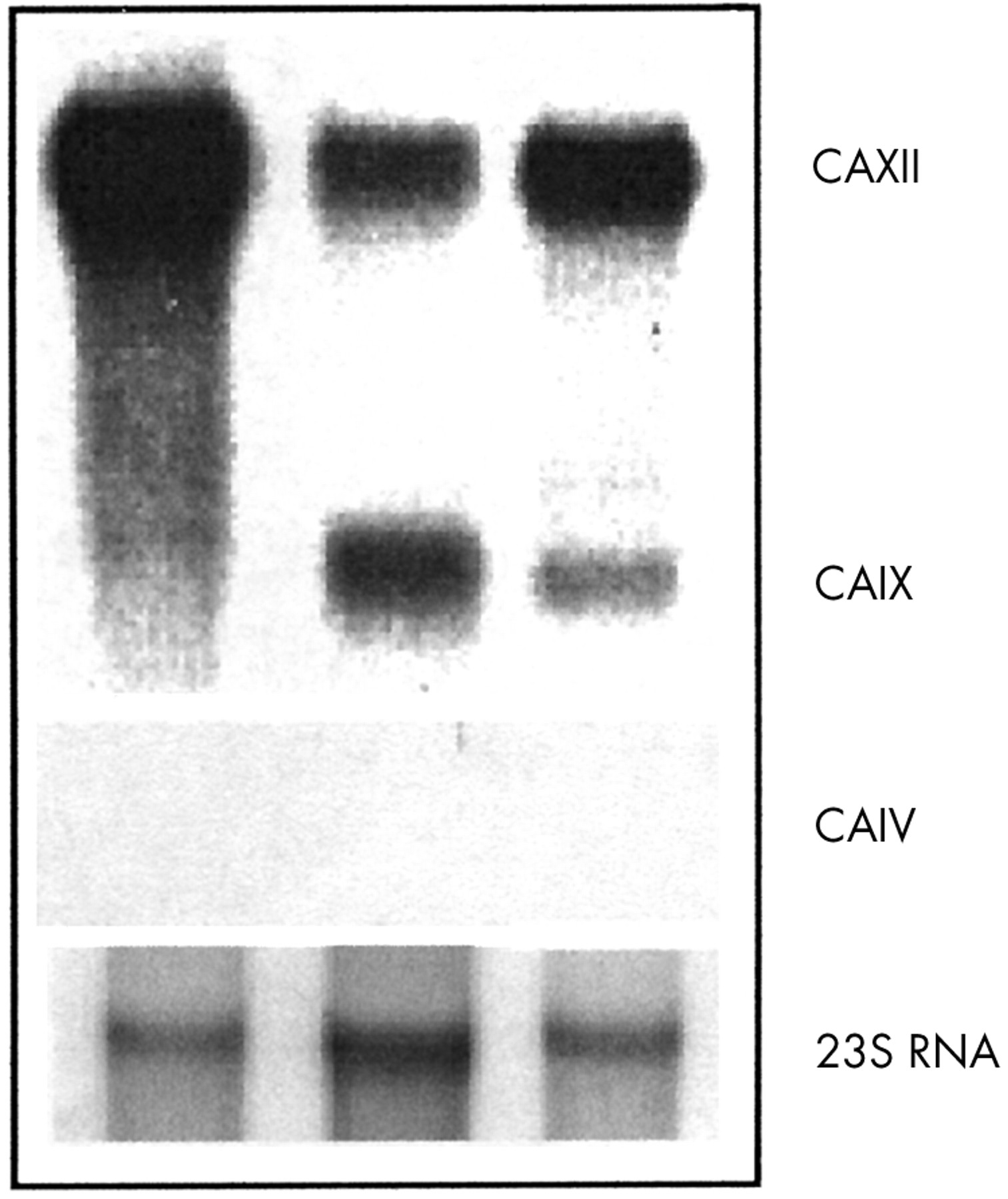
We then examined the expression of these genes by northern blot analysis of mRNA isolated from these, normal (2 year old), and glaucomatous ciliary NPE cells grown to confluence (fig 2). They showed relatively strong CA9 and CA12 signals on northern blots from a normal subject while, in contrast, NPE cells from a glaucoma patient showed high (five times) overexpression levels of the CA12 mRNA with no detectable expression of CA9, consistent with the immunostaining data (table 1). In this experiment we also confirmed the absence of expression of the CA4 gene as was shown previously by immunostaining of eye sections with specific CAIV antibodies.3 Thus, the assumed involvement of this enzyme in the ciliary epithelium7,8 was not corroborated by our experiments.
Northern blot analysis of CA9, CA12, and CA4 expression in cultures of human ciliary non-pigmented epithelial cell lines from a glaucoma patient (GCE-T, passage 12), a normal subject (ODM-C4, passage 9), and, as a positive control, from the renal carcinoma cell line 786-0.12 28S rRNA levels indicate equivalent loading of mRNA in all lanes. As expected, strong positive hybridisation with the CA4 probe (~1.35 kb band) was obtained with many human tissues on MTN 7760-1(Clonetech, Palo Alto, CA, USA) northern blots (data not shown).
In the present work, we have established the nature of the CAs expressed and most likely involved in the development and function of the ciliary epithelium and shed new light on the molecular pathogenesis of glaucoma. Glaucoma is a complex, relatively common (affects over 67 million people world wide2) eye disorder, characterised by clinical and genetic heterogeneity.2,16,20 The mode of transmission is complex and involves multiple causative and modifying susceptibility genes. To date, at least 10 loci that potentially confer susceptibility to glaucoma have been mapped and so far only two causative candidate genes have been identified.17–19 Previously, it was assumed by many working on the aqueous humour production that CAIV is the crucial membrane enzyme involved in the ciliary process.7,8 Contrary to this belief, we establish here that the novel cell surface bitopic transmembrane enzymes CAIX and CAXII are expressed in the ciliary cells and most likely play an important role in aqueous humour production. Classical theories of glaucoma have focused primarily on the assumption that faulty facilities governing the outflow system may underlie the physical and genetic causes of this multifaceted disease.20 Here, we show for the first time the overexpression of the CAXII enzyme in the ciliary NPE cells of glaucoma patients, suggesting that some glaucomas might result from overproduction of the aqueous humour solely or in combination with damage to the outflow facility. The silencing of the CA9 gene in adult eyes (probably by hypermethylation21) could lead in some people to overexpression of the CA12 gene in the ciliary NPE cells, which may in turn cause overproduction of the aqueous humour and subsequently high intraocular pressure and hence lead to glaucoma. Furthermore, it is also possible that the overexpression of CAXII in glaucoma patients may be caused by mutated allele(s) of this gene. Clearly, the overexpression of CA12 could be a diagnostic marker for certain types of glaucomatous ciliary NPE cells and may provide a framework for better understanding of the fluid equilibrium in the eye. In clinical terms it should also impact on the quest for more selective topical inhibitors of CAXII for the treatment of glaucoma. We have recently identified novel sulphonamide inhibitors selectively inhibiting CAXII or CAIX using purified recombinant CAXII and CAIX enzymes (F Jurnak et al, in preparation).
Acknowledgments
This work is dedicated to the memory of the late Professor Thomas Maren, a pioneer in carbonic anhydrase research and development of topical carbonic anhydrase inhibitors for the treatment of glaucoma. We wish to thank Professor William S Sly for providing the CAXII antibody. This work was supported in part with federal funds from the National Cancer Institute, National Institutes of Health, under Contract No NO1-CO-56000, No NO1-CO-12400, by NCI grant CA19401, and NIH/NEI grant EY04873. The content of this publication does not necessarily reflect the views or policies of the Department of Health and Human Services, nor does mention of trade names, commercial products, or organisations imply endorsement by the US Government.