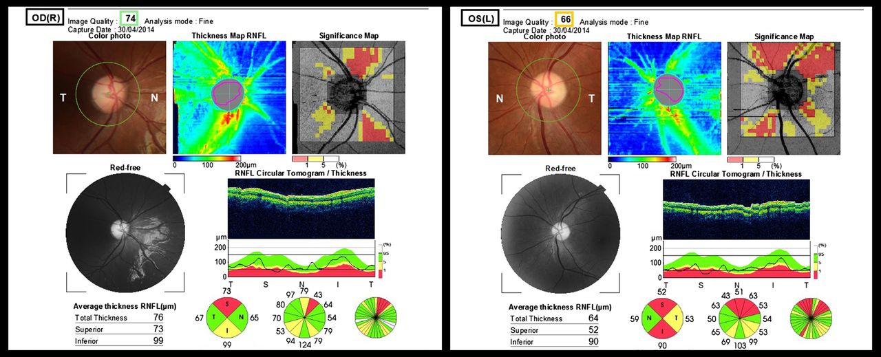
Topcon optical coherence tomography (OCT) of the retinal nerve fibre layer and fundal pictures of an 11-year-old female (case no. 8 in the table) with methylmalonic acidemia. Diagnosed as a neonate, previous complications included severe renal failure and extrapyramidal movement disorders following earlier basal ganglia infarction. She presented with subacute bilateral decrease of visual acuity recorded at 6/60 for right and left eyes and bilateral dyschromatopsia, noted concomitant to a severe episode of metabolic decompensation with initial normal fundoscopy and mildly subnormal acuities. Four months later, fundal pictures show bilateral disc pallor with asymmetric retinal nerve fibre layer thinning of 76 μm in the right eye and 64 μm in the left on OCT. At the time of the OCT, visual acuity had improved and was 6/9.5 and 6/30 for right and left eyes, respectively.


Share this article
Click the icon of the social media platform on which you would like to share this article.


