
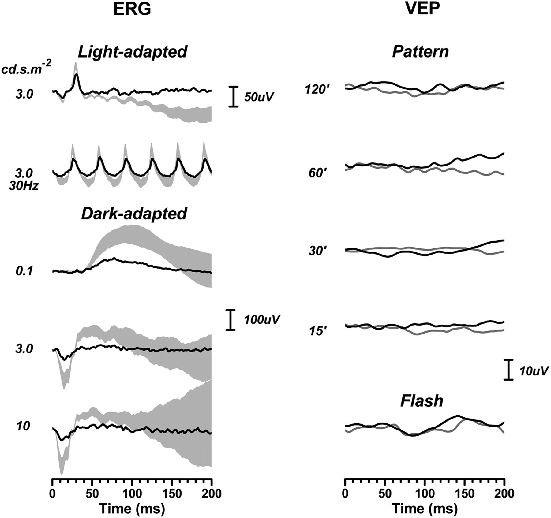
Electrodiagnostics, from case 12, showing absent flash and pattern flash visual evoked potentials (VEPs) (right column). Electroretinogram (ERGs) were recorded in cases 6, 7 and 12 using corneal fibre electrodes and complied with international standards. Case 10 had ERGs recorded using skin electrodes, for reasons of compliance. Only case 12 is shown because the findings from the three other patients are remarkably similar. The light-adapted (cone) responses are attenuated to some extent but this is much greater in the dark-adapted state, suggesting a rod-mediated retinal dysfunction. The shaded areas in the ERG depict 95% CIs from our normative data set. The VEP data show 2 responses per condition.


Share this article
Click the icon of the social media platform on which you would like to share this article.


