Article info


- Dr Giuseppe Querques, Department of Ophthalmology, University of Paris XII, Centre Hospitalier Intercommunal de Creteil, 40 Avenue de Verdun, 94000 Creteil, France; giuseppe.querques{at}hotmail.it
Citation
Publication history
- First published November 16, 2007.
Video Report
A new approach for visualisation of dye leakage in fluorescein angiography
Giuseppe Querques (1,2), Nicola Delle Noci (2), Gisele Soubrane (1), Eric H Souied (1)1 Department of Ophthalmology, University of Paris XII, Centre Hospitalier Intercommunal de Creteil, 40 Avenue de Verdun, 94000 Creteil, France. 2 Department of Ophthalmology, Policlinico Ospedali Riuniti, University of Foggia, Italy.
Correspondence: Dr Giuseppe Querques, Email: giuseppe.querques{at}hotmail.it Department of Ophthalmology, University of Paris XII, Centre Hospitalier Intercommunal de Creteil, 40 Avenue de Verdun, 94000 Creteil, France. Tel: +33 (0)1 45 17 52 22; Fax: +33 (0)1 45 17 52 66.
Date of acceptance: 30th September 2007
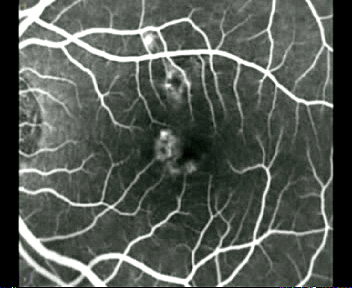
Video 1. Fluorescein angiography movie of an active leaking newly diagnosed extrafoveal classic choroidal neovascularization.
Video 2. Fluorescein angiography movie of an active leaking newly diagnosed subfoveal minimally classic choroidal neovascularization.
Video 3. Fluorescein angiography movie of a juxtafoveal classic choroidal neovascularization previously treated by photodynamic therapy: fibro-atrophy temporally, minimal active leakage nasally.
Video 4. Fluorescein angiography movie of a subfoveal occult choroidal neovascularization previously treated by photodynamic therapy (PDT): filling of the vascularized pigment epithelium detachment.
Video 5. Fluorescein angiography movie of an active central serous chorioretinopathy: visualization of the kinetics of the classic smokestack-type leakage.
Video 6. Fluorescein angiography movie of an active chronic central serous chorioretinopathy: visualization of dye leakage from recurrent active areas of central serous chorioretinopathy.View Video: Fast connectionView Video: Dial up connection
Note: This video is best viewed in Quicktime
Introduction
Fluorescein Angiography (FA) is the more common investigation performed for macular diseases.(1) Frozen FA pictures are obtained but direct visualization of the kinetics of FA is possible only once, by the investigator. The kinetics of FA examination is imagined from static FA pictures based on our experience.(2,3) We evaluated a new software that re-create automatic pseudo-movie from static pictures, in order to share the visualization of dye leakage.
Methods
EyeToolkit software (EDC Lamy EyeToolkit software®, EDC LAMY, Carvin, France) performs an automatic and rapid overlay (0.5 to 3 seconds) from the different angiographic frames. Five images from the same examination, from early to late phase, are required to generate a movie. The resulting movies could be seen either with the EyeToolkit software or be exported to video format.
We submitted a minimum of 6 images issued from printed photographs or any digital instrument (Topcon retinal camera, TRC-50, Topcon, Tokyo, Japan; Heidelberg Retina Angiograph , HRA 2, Heidelberg Engineering, Heidelberg, Germany), to the EyeToolkit software: sequences of fluorescein angiograms of patients harbouring various patterns of exudative age-related macular degeneration (AMD), with or without treatment, were selected in order to visualise active leakage of dye and to differentiate it from staining.
In addition, we submitted to the EyeToolkit software images issued from one single FA examination (early phase to 10’) in case of central serous chorioretinopathy (CSC), in order to see the kinetics of the leakage and to visualise leakage of dye from active areas of CSC.
Comment
We evaluated two kind of leakage: chorio-retinal leakage from choroidal newvessels (CNVs) due to AMD, and retinal leakage from active areas of CSC.
In exudative AMD patients, the rapid overlay (<10’’) applied in a movie mode obviously demonstrated the progressive enlargement of the hyperfluorescence, materializing the leakage of fluorescein from CNVs. The FA movies generated with EyeToolkit permitted to clearly see the active leakage from a newly diagnosed classic CNV (Video 1), and from a newly diagnosed minimally classic CNV (Video 2). Moreover, the FA movies generated with EyeToolkit permitted to easily detect the minimal leakage from a still active classic choroidal neovascularization previously treated by photodynamic therapy (PDT) (Video 3), and to visualise the filling of the vascularized pigment epithelium detachment from an occult choroidal neovascularization previously treated by PDT (Video 4).
In CSC, the FA movies generated with EyeToolkit permitted to see the kinetics of the classic smokestack-type leakage (Video 5) and to easy visualise leakage of dye from active areas of CSC (Video 6).
EyeToolkit software is a new automatic and efficient tool for a rapid overlay of images, obtaining an angiographic movie from any images. It leads to easy visualisation of leakage from choroidal newvessels and from active areas of CSC; thus, owing to the easy differentiation of active leakage from staining, EyeToolkit software would seem particularly helpful for the decision of treatment and re-treatment. Easy visibility of the kinetics of the dye in angiography, in a movie mode, also represents a useful approach for teaching.
Based on this preliminary analysis, this software can be considered for:
(a) Recreating pseudo-kinetics of dye in retinal angiography;
(b) Easy sharing of retinal angiograhy; voice comments can be included within the file;
(c) Teaching and e-learning.
The comparative evaluation of the generated movies versus classic angiographic pictures must validate its use for diagnosis and indications of treatment.Conflict of Interest Statement
The authors have no financial interest in the software product used in this study.
References
- Gass JMD. Stereoscopic altas of macular diseases diagnosis and treatement. Saint-Louis, 1987, The CV Mosby Co, 3 ed.
- Amalric P. New developments in fluorescein angiography. Doc Ophthalmol 1977 29;43:127-135.
- Mainster MA, Timberlake GT, Webb RH, Hughes GW. Scanning laser ophthalmoscopy. Clinical applications. Ophthalmology 1982 Jul;89:852-7.
Files in this Data Supplement:
Request permissions
If you wish to reuse any or all of this article please use the link below which will take you to the Copyright Clearance Center’s RightsLink service. You will be able to get a quick price and instant permission to reuse the content in many different ways.