Article Text
Abstract
Introduction: Post operative complications following cataract surgery can present following a seemingly normal procedure, with few clues as to cause.
Case Description: A 62-year-old female had cataract surgery on her left eye. The surgery was uncomplicated until the end of the procedure. When the surgeon inserted a small cannula with balanced salt solution, to refill the anterior chamber just prior to checking the wound, the cornea gradually turned somewhat translucent. The following morning the cornea was cloudy and the visual acuity was count fingers.
The patient was referred and found to have visual acuity of count fingers without pain and normal intraocular pressure. An Ultrasound Biomicroscopic Examination (UBM) was performed, revealing a detached Descemet’s membrane, which could be seen in the anterior chamber waving within the aqueous. Five days later, reattachment of Descemet’s membrane was performed with air without complication. Within 4 weeks, the visual acuity was 6/12 with retinal pathology accounting for the remaining reduction in visual acuity.
Discussion: This patient was referred with a diagnosis of endothelial decompensation. The surgeon assumed that he had injected a toxic solution into the anterior chamber. The use of UBM allowed the diagnosis of Descemet’s detachment to be made in the setting of an opacified cornea. Repair with the use of air to re-approximate the membrane with its complement of endothelium led to visual recovery in four weeks.
Statistics from Altmetric.com
Supplementary materials
Video Report
Detached Descemet's membrane
Ivan R Schwab, Ellen RedenboUniversity of California, Davis Department of Ophthalmology & Vision Science, California
Correspondence: Dr Ivan R Schwab
Email: irschwab{at}ucdavis.edu Department of Ophthalmology & Vision Science, UC Davis Health System, 4860 Y Street, Suite 2400, Sacramento, CA 95817, USADate of acceptance: 7th June 2007

A 62 year-old woman presented with a cloudy cornea and count fingers vision following cataract surgery. An Ultrasound Biomicroscopic examination (UBM) revealed a detachment of Descemet's membrane, which can be seen waving within the aqueous. View Video: Fast connectionView Video: Dial up connection
Note: This video is best viewed in Quicktime
Introduction
Post operative complications following cataract surgery can present following a seemingly normal procedure, with few clues as to cause.
Case Description
A 62 year old female had cataract surgery on her left eye. The surgery was uncomplicated until the end of the procedure. The surgeon had inserted the lens into the capsular bag, and removed the viscoelastic from the anterior chamber. The surgeon inserted a small cannula with balanced salt solution to refill the anterior chamber just prior to checking the wound. The cornea gradually turned somewhat translucent. The surgeon checked the wound and found it be water tight, and applied a patch. The following morning the cornea was cloudy and the visual acuity was count fingers.
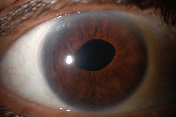
The patient was referred to our office and was found to have visual acuity of count fingers without pain and normal intraocular pressure. An Ultrasound Biomicroscopic Examination (UBM) was performed and is attached. A diagnosis of a detached Descemet's membrane was made. Five days later, reattachment of Descemet’s was performed with air without complication. Within four weeks, the visual acuity was 6/12 with retinal pathology accounting for the remaining reduction in visual acuity. A photograph of postoperative appearance at three weeks is shown in Figure 1.
Discussion
This patient was referred with a diagnosis of endothelial decompensation. The surgeon assumed that he had injected a toxic solution into the anterior chamber. Visualization of the anterior chamber was difficult, but the UBM confirmed the detachment of Descemet's membrane as can be seen from the video. Note Descemet's waving in the aqueous. Repair was straight forward with the use of air to re-approximate the membrane with its complement of endothelium. Visual recovery required approximately four weeks.

Figure 1 Slit lamp photograph showing the appearance 3 weeks after successful reattachment of Descemet’s membrane.Files in this Data Supplement:
Footnotes
To view the full report and accompanying video please go to: http://bjo.bmj.com/cgi/content/full/91/9/1132/DC1
All videos from the BJO video report collection are available from: http://bjo.bmj.com/video/collection.dtl


