Article Text

ABSTRACT
Fourier-domain optical coherence tomography (OCT) was used to image the three-dimensional (3D) structures of the proliferative membrane in proliferative diabetic retinopathy. The case of a 51-year-old man with retinal detachment of the macula in his left eye is reported. The proliferative membrane covered the entire macular area. In the OCT image, the 3D structure of the proliferative membrane could be clearly visualised. The OCT image showed the presence of multiple adhesions between the retina and the proliferative membrane and separation of the proliferative membrane. The patient underwent three-port vitrectomy, and the extent and locations of the adhesions corresponded well with the findings during vitrectomy. Three-dimensional OCT is an effective tool for understanding the 3D structure of the proliferative membrane in diabetic retinopathy and is useful for training and planning of the surgical procedures in vitrectomy.
To view the full report and accompanying video please go to: http://bjo.bmj.com/cgi/content/full/92/5/713/DC1
All videos from the BJO video report collection are available from: http://bjo.bmj.com/video/collection.dtl
Statistics from Altmetric.com
Supplementary materials
Video Report
Three-dimensional optical coherence tomography of proliferative diabetic retinopathy
T Iwasaki(1,3), M Miura1(2,3), C Matsushima1(3), M Yamanari(2,3), S Makita(2,3), Y Yasuno(2,3)1 Department of Ophthalmology, Tokyo Medical University, Kasumigaura Hospital, Inashiki, Ibaraki, Japan; 2 Computational Optics Group in the University of Tsukuba, Tsukuba, Ibaraki, Japan; 3 Computational Optics and Ophthalmology Group Correspondence to:
Correspondence: Dr M Miura
Email: m-miura{at}tokyo-med.ac.jp Department of Ophthalmology, Tokyo Medical University, Kasumigaura Hospital, 3-20-1 Chuo, Ami, Inashiki, Ibaraki 3000395, Japan. Tel: +81-298-87-1161; Fax: +81-298-87-7656.Date of acceptance: 4th March 2008
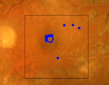
Figure 1 Color fundus photograph showing the proliferative membrane. The black lines indicate the areas of three-dimensional optical coherence tomography imaging. Blue indicates the adhesions between the retina and the proliferative membrane.
Video 1 Series of B-scan optical coherence tomography (OCT) images, ranging from superior to inferior. B-scan OCT images showing multiple adhesions between the retina and the proliferative membrane. Separation of the proliferative membrane and the macular hole are observed.
Video 2 Three-dimensional OCT image. Three-dimensional OCT image showing the three-dimensional structure of proliferative membrane and the presence of multiple adhesions.View Video: Fast connectionView Video: Dial up connection
Note: This video is best viewed in Quicktime
Introduction
Proliferative membrane formation in diabetic retinopathy leads to various complications such as vitreous hemorrhage and tractional retinal detachment. These complications are important causes of vision deterioration in diabetic retinopathy. Vitrectomy with membrane resection is the most common method for treating the proliferative membranes. For a safe and successful surgical procedure, an understanding of the three-dimensional structure of the proliferative membrane and underlying retina is crucial. During conventional fundus examination, evaluation of the structures beneath the proliferative membrane is frequently difficult and complete three-dimensional information regarding the proliferative membrane can be rarely obtained. Fragmentary information about the proliferative membrane and underlying retina can be obtained with the B-Scan images of the time-domain optical coherence angiography (OCT);1 however, the entire structure of the proliferative membrane can rarely be detected. Recently, dramatic advances in the OCT technology have enabled three-dimensional OCT imaging.2,3 Fourier-domain OCT allows imaging with dramatically higher imaging speeds than those provided by a conventional time-domain OCT system. The measurement speed in the Fourier-domain OCT is higher because of the absence of mechanical scanning (A-scan). Using Fourier-domain OCT, three-dimensional imaging of the proliferative membrane could be possible. In this video report, we evaluated the three-dimensional structure of the proliferative membrane in diabetic retinopathy using Fourier-domain OCT and correlated these findings with the surgical procedure of vitrectomy.
Observation
A 51-year-old man presented with an 8-month history of progressive vision loss in the left eye. Best-corrected visual acuity in his left eye was 10/200. Conventional fundus examination showed proliferative diabetic retinopathy with retinal detachment of the macula in his left eye. Contact lens biomicroscopy of the left eye showed the presence of the proliferative membrane covering the entire macular area; however, the three-dimensional structures underlying the proliferative membrane were hardly observed. A three-dimensional OCT image was obtained using the commercialized Fourier-domain OCT (Topcon 3D-1000, Topcon, Tokyo, Japan). The approximate measurement area was 6 mm × 6 mm on the retina, and a raster-scanning protocol with 256 A-scans × 256 B-scans was used. The scanning rate was 18000 A scans/s, and the acquisition time for the three-dimensional image was 3.6 s. The video is regarding three-dimensional OCT imaging of the proliferative membrane. A series of B-scan OCT images demonstrated multiple adhesions between the retina and the proliferative membrane. At the fovea, the proliferative membrane pulled the retina tangentially and induced a macular hole. The proliferative membranes were perpendicularly separated at the upper and lower regions to the macula. At the upper region, the B-scan OCT images showed individual adhesions of the inner and outer proliferative membranes to the retina. The three-dimensional OCT images revealed the three-dimensional structure of the proliferative membrane and the presence of multiple adhesions. At each adhesion, the detached retina was pulled toward the proliferative membrane, thus implying that imprudent surgical intervention of the adhesions might cause iatrogenic retinal breaks. At the fovea, the adhesion had encircled the macular hole to some extent. In other areas, the adhesions were located adjacent to the retinal vessels and were classified as epicenters. At the epicenters, the retina was attached to the proliferative membrane in point-like areas. Perpendicular separation of the proliferative membranes was observed in the upper and lower regions to the macula. From the three-dimensional OCT data set, we detected the adhesions using an image processing software (Amira 4.1, Mercury Computer Systems, Chelmsford, MA, USA). After detecting the adhesions in the three-dimensional OCT images, the extent and the locations of the adhesions were registered in the color fundus photograph (fig 1). The patient underwent 25-gauge three-port vitrectomy. The three-dimensional OCT information corresponded well with the findings during vitrectomy, and this information proved valuable for safe and successful surgical resection of the membrane. After successful vitrectomy, the entire retina was attached, and the visual acuity improved to 20/200.
Comment
In this video report, we evaluated the three-dimensional OCT imaging of proliferative diabetic retinopathy and assessed the anatomic morphology of the proliferative membrane and the accompanying multiple adhesions. Fourier-domain OCT provided information regarding the complete three-dimensional structure of the proliferative membrane that could be hardly obtained with conventional fundus examination or time-domain OCT. The three-dimensional information provided by Fourier-domain OCT has potential applications for vitreous surgery. Understanding these three-dimensional structures is frequently difficult for inexperienced and intermediate-level surgeons being trained in vitrectomy. Understanding anatomic morphology could be easier if three-dimensional OCT images are used, and thus it could prove very useful for the education and training regarding vitrectomy. For skillful vitreous surgeons, the three-dimensional information such as location of the epicenters is beneficial for safe and successful surgical resection of the membrane. Thus, three-dimensional OCT is an effective tool for understanding the three-dimensional structure of proliferative membrane in proliferative diabetic retinopathy.
Funding and competing interests
Supported in part by the Grant-in-aid for Scientific Research 15760026 from the Japan Society for the Promotion of Science, Japan Science and Technology Agency, and the Special Research Project of Nanoscience at the University of Tsukuba.
Each author states that he or she has no proprietary interest in the development or marketing of this or a competing instrument or software.
References
- Gallemore RP, Jumper JM, McCuen BW, 2nd, et al. Diagnosis of vitreoretinal adhesions in macular disease with optical coherence tomography. Retina. 2000;20:115-120.
- Wojtkowski M, Bajraszewski T, Targowski P, et al. Real-time in vivo imaging by high-speed spectral optical coherence tomography. Optics Letters 2003;28:1745-1747.
- Schmidt-Erfurth U, Leitgeb RA, Michels S, et al. Three-dimensional ultrahigh-resolution optical coherence tomography of macular diseases. Invest Ophthalmol Vis Sci. 2005;46:3393-3402.
Files in this Data Supplement:
Footnotes
Funding: Supported in part by the Grant-in-aid for Scientific Research 15760026 from the Japan Society for the Promotion of Science, Japan Science and Technology Agency, and the Special Research Project of Nanoscience at the University of Tsukuba.
Competing interests: None.
TI and MM contributed equally to this study.