Article Text

Abstract
Purpose We define the ideal anterior capsulotomy through consideration of capsular histology and biomechanics. Desirable qualities include preventing posterior capsular opacification (PCO), maintaining effective lens position (ELP) and optimising capsular strength.
Methods Laboratory study of capsular biomechanics and literature review of histology and published clinical results.
Results Parameters of ideal capsulotomy construction include complete overlap of the intraocular lens to prevent PCO, centration on the clinical approximation of the optical axis of the lens to ensure concentricity with the capsule equator, and maximal capsular thickness at the capsulotomy edge to maintain integrity.
Conclusions Constructing the capsulotomy centred on the clinical approximation of the optical axis of the lens with diameter 5.25 mm optimises prevention of PCO, consistency of ELP and capsular strength.
- Treatment Lasers
This is an Open Access article distributed in accordance with the Creative Commons Attribution Non Commercial (CC BY-NC 4.0) license, which permits others to distribute, remix, adapt, build upon this work non-commercially, and license their derivative works on different terms, provided the original work is properly cited and the use is non-commercial. See: http://creativecommons.org/licenses/by-nc/4.0/
Statistics from Altmetric.com
Creating the capsulorhexis is often acknowledged as both the most challenging and critical step in a cataract procedure.1 ,2 A properly constructed capsulorhexis provides the foundation for lens extraction and stable in-the-bag intraocular lens (IOL) fixation. A perfectly circular and properly sized capsulorhexis allows the capsular bag to completely envelop the IOL optic, reducing the incidence of posterior capsular opacification (PCO) and providing a more predictable effective lens position (ELP). The capsular opening created requires mechanical strength sufficient to ensure the integrity of the capsule during lens extraction and IOL implantation.
In order to ensure that the anterior capsule overlaps the IOL edge 360° after implantation, a manually created continuous curvilinear capsulorhexis (CCC) must be created with an average diameter somewhat smaller than ideal because of unavoidable variation in centration, diameter and circularity.3 With femtosecond laser capsulotomy, precise placement of the capsulotomy with significantly less unintended variation in location and diameter is possible.4 Achieved capsulotomy diameters have been shown to be very close to the intended measurements.5 Therefore, given increased precision in capsulotomy construction, the question arises as to the ideal location and size of the capsulotomy.
Several considerations bear on the discussion of ideal capsulotomy design. These include preventing PCO, maintaining consistent ELP and maximising mechanical strength.
Prevention of PCO
Enhancement of the barrier effect to lens epithelial cell growth through complete overlap of the IOL optic by the anterior capsule edge has become well established in clinical practice. Ravalico et al6 suggested in 1996 that a ‘capsulorhexis with a slightly smaller diameter than the IOL optic appears to be better than a large-size capsulorhexis in reducing the incidence of PCO.’ The study by Hollick et al7 confirmed significantly less PCO with a capsulorhexis completely covering the edge of the IOL optic. Apple et al stated, “Our histopathological observations suggest that creating a [continuous curvilinear capsulorhexis] with a diameter slightly smaller than that of the IOL optic allows the capsule edge to adhere to the anterior surface of the optic, enhancing the efficiency of the barrier effect by creating a closed system.”8
More recently, Kovacs et al have suggested that femtosecond laser capsulotomies reduce the incidence of PCO due to superior IOL positioning. In a comparative study of manual capsulorhexis and femtosecond laser capsulotomy, they found that vertical tilt, horizontal and total decentration of IOLs, and PCO proved to be significantly higher in the manual capsulorhexis group (p=0.03, 0.04, 0.03 and 0.01, respectively). After adjusting for axial length and follow-up time, manual anterior capsulorhexis represented ‘a significant predictor of higher PCO scores in the multivariable regression model (β: 0.33; 95% CI 0.01 to 0.65; p=0.04).’9 These authors concluded that improved IOL positioning led to a reduced PCO in the cases performed with the femtosecond laser.
The evidence to date supports a capsulotomy edge that completely overlaps and adheres to the IOL optic, and thus defines the first parameter of the ideal capsulotomy: 360° optic overlap. This requirement applies equally to manual capsulorhexis and laser capsulotomy: it is the accurate, reproducible placement of the laser capsulotomy that may primarily convey an advantage in this regard. Given the dimensions of the vast majority of IOLs implanted today, this requirement translates into an actual physical capsulotomy diameter <6.0 mm. Surgeons should, of course, be aware of the apparent magnification of the capsulotomy when observed through the cornea.
Maintaining consistent ELP
Inaccuracies in the determination of the ELP represent a very important factor in determining the effective power of an IOL in the eye.10 For example, it has been previously calculated that a 1.4 D spherical refractive error results from forward displacement of a 21 D IOL (calculated to produce postoperative emmetropia) by 1 mm.11 Several studies, as described below, have suggested a relationship between capsulorhexis or capsulotomy design and ELP. Standardisation of capsulotomy construction may therefore permit more consistent outcomes.
Cekiç and Batman12 demonstrated that a capsulorhexis diameter of 4.0 mm resulted in a longer postoperative anterior chamber depth (ACD) than a 6.0 mm capsulorhexis. Their paper sparked initial interest in the possibility of controlling ELP through capsulorhexis design.
Published studies of femtosecond laser capsulotomy have shown an improvement in refractive outcomes. Kránitz et al13 demonstrated in a prospective, randomised, controlled trial that a laser capsulotomy resulted in a more stable refractive result when compared with a more variable manual capsulorhexis. In addition, Hill et al showed a reduction in postoperative mean absolute error (MAE) from 0.59±0.35 D in a group of 123 cases with a manual capsulorhexis to 0.42±0.39 D in a matched group of 249 cases with a femtosecond laser capsulotomy (p<0.001). A total of 47.4% of the laser treated eyes were within 0.25 D of intended postoperative manifest spherical equivalent refraction, compared with 22.0% for the manual eyes (p=0.003).14 Filkorn et al likewise demonstrated significantly lower MAE in 77 eyes of 77 patients undergoing laser capsulotomy (0.38±0.28 D) than in 57 eyes of 57 patients undergoing manual capsulorhexis (0.50±0.38 D; p=0.04). The difference was greatest in short (axial length <22.0 mm, 0.43±0.41 vs 0.63±0.48) and long (axial length >26.0 mm, 0.33±0.24 vs 0.63±0.42) eyes.15
On the other hand, studies of manual capsulorhexis have not consistently shown an effect of size and centration on postoperative ELP. Findl has suggested that capsulorhexis size and shape do not have a significant influence on postoperative IOL tilt, decentration or ACD, even if the capsulorhexis is large or eccentric.16 Davidorf similarly found that there is no relationship between capsulorhexis morphology, lens decentration and tilt and refractive outcomes in cataract surgery.3 Davison found that ‘imperfection of optic overlap had no anatomic or refractive clinical significance.’17
Taken at face value, the apparent contradiction between these groups of studies, some of laser capsulotomy and others of manual capsulorhexis, may actually reflect the greater variability inherent in manual capsulorhexis construction compared with capsulotomies constructed by the femtosecond laser. For example, using manual capsulorhexis, Findl reported SDs for mean IOL tilt and decentration of ±2.1° and ±0.2 mm, respectively.12 Kranitz et al, using femtosecond laser surgery, reported SDs for tilt of ±1.08° (horizontal) and ±1.41° (vertical); they reported an SD for total decentration of ±111.54 μm. These data show that the variance of IOL tilt is 2–4 times greater with manual capsulorhexis than it is with laser capsulotomy; the variance of IOL decentration is about four times greater. It is, of course, more difficult to show a statistically or clinically significant difference within or among groups of data when each group has a high degree of internal variance. Attempts to develop intraocular instrumentation to improve the construction of the manual capsulorhexis demonstrate the perceived need for greater accuracy.18 ,19 Studies of laser capsulotomy have demonstrated improvements in IOL positioning and reduction of postoperative refractive error.
Reduction of systematic errors depends on use of consistent methodology and measurement of outcomes. By sizing and locating the capsulotomy in a reproducible and consistent fashion, the femtosecond laser removes a significant amount of variability from the cataract extraction system. Removing this variability allows constant improvement through ongoing outcomes analysis and optimisation of the ‘A constant’ or its equivalent in IOL power calculation formulae.
Maximising mechanical strength
Recently, Abell et al20 have suggested a relatively higher incidence of anterior capsule tears with femtosecond laser cataract surgery. Other case series have observed no difference,21 or the effect of a learning curve, with significant reduction of complication rates over time.22 Worldwide experience indicates a very low rate of anterior capsule tears after femtosecond laser assisted capsulotomy.23
While improvement in surgical technique undoubtedly plays a role in the learning curve phenomenon,24 the parameters of laser capsulotomy may also have an impact on safety and effectiveness. For example, in Abell's report, the mean capsulotomy diameter is noted to have always been <5.0 mm, and ‘typically’ 4.7 mm.19 A review of lens capsule anatomy suggests that a somewhat larger capsulotomy would provide a superior safety profile.
Histology shows that the anterior human lens capsule is not of uniform thickness. Barraquer et al measured human capsules using digital micrographs and found that mean capsular thickness increased with age from 11 to 15 µ at the anterior pole and from 13.5 to 16 µ at the anterior midperiphery. They found a local thinning at the pre-equatorial zone, which changed little with age. The equatorial thickness remained constant at 7 µ. At the posterior periphery, thickness decreased with age from 9 to 4 µ. There was no change in thickness at the posterior pole (overall mean, 3.5 µ).25 This variation in thickness is thought to derive in part from the anchoring of zonular fibres in the peripheral capsule and from development of the capsule for the role that the capsule plays in the mechanism of accommodation.26 As shown in figure 1, using the value of 9.8 mm for average lens diameter, the peak thickness occurs at a diameter from 4.9 to 5.5 mm, centred on the anterior pole.27 These histological studies suggest that a capsulotomy of 5.25 mm, centred on the anterior pole of the lens capsule, would be most likely to intersect the anterior capsule at its thickest (and therefore strongest) point.

Diagram of the capsule after Fincham, 1937,40 scaled to an equatorial diameter of 9.8 mm with one half of the capsule divided into 200 divisions in 10 division steps.25 The thickest part of the capsule, in accordance with the results from Barraquer, is between divisions 40 and 50 and occurs at a diameter of approximately 5 mm or greater.
Additionally, the ability of the capsule edge to resist tearing increases with the diameter of the capsulotomy. To elucidate this phenomenon, we investigated the effect of capsulotomy diameter from 4.0 to 5.5 mm on extensibility and break force in porcine eyes.28 Because of anatomic differences and the higher elasticity of porcine lens capsules, the results obtained in this study may not be fully comparable with those in a clinical setting. Nevertheless, the porcine capsule has been validated as a model for the paediatric human eye,29 and it has proved useful in prior studies of femtosecond laser capsulotomy.30 Performing the same measurements in human cadaver eyes is possible, but limitations do exist, including elapsed time since enucleation, transportation and the age of the donor.31 Maximal capsular stretch in the human has been noted to decrease significantly with age, although capsular elasticity has not demonstrated a similar variation.32
Methods
To investigate the variation of capsule resistance to tearing with capsulotomy diameter, a total of 49 freshly enucleated porcine eye lens capsules were tested for tensile strength. All eyes were obtained from a local processing plant and maintained at room temperature in balanced salt solution (BSS) prior to use. The porcine eyes were randomly assigned to the following treatment groups: 14 eyes were assigned to the manual CCC group and 35 eyes were assigned to one of the laser capsulotomy (Laser 1–4) groups as shown in table 1.
Input parameters for the laser capsulotomy treatment
The Laser eyes were divided into four subgroups (Laser 1–4) based on the capsulotomy diameter and laser parameters.
We defined the capsulotomy break force as the force measured when the capsule ruptures; the maximum extensibility measured how far the capsular opening was stretched beyond the zero point before breaking. Stretching of the lens capsules to ascertain the elongation and force at fracture was performed in each group using a custom set of pin arms designed to hold the lens capsule passively. Once attached to the measuring arms, the unstretched diameter of the capsular opening was measured using callipers. With this experimental set up, stretching of the capsule occurred in a manner that provided a uniaxial vertical load without any horizontal or torsional forces. The stretching apparatus consisted of two plastic stretching pins (thermoplastic polysulfone), circular in cross section and 2 mm in diameter, one of which gradually moved apart in a translational motion from the second, pulling on the capsular rim while the stretching force was measured. The pins were oriented horizontally and the globes were maintained laterally on a metallic L-shaped seat attached to a cantilever that could be raised and lowered to apply gentle contact of the capsular rim and the fixed probe. The system apparatus is shown in figure 2.

Testing apparatus.
The two probe pins were inserted into the capsulotomy opening. One probe was fixed and connected to a load cell (Acculab–VIC 123) for force measurement, and the second probe was attached to a stepper motor (850G Actuator), programmed to travel at a velocity of 62 μm/s in 500 μm intervals with 3 s pauses to allow data acquisition. Custom computer software controlled the motor while recording the data from the force transducer. For both CCC and Laser tests, the eyes were resting on a cantilever seat and lowered into a BSS contained in a small lab cuvette. The globes were maintained deliberately immersed in the BSS during the test to prevent drying and osmotic changes.33
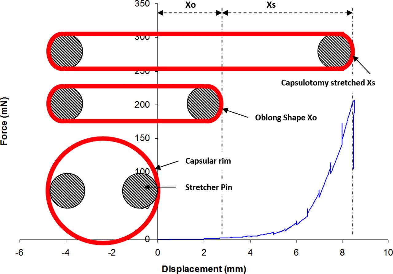
The stretching experiments were composed of three successive phases as presented in figure 3. The first phase shown in the bottom diagram in figure 3 corresponds to the pin arms positioned to be in contact with the capsular rim at 6 and 12 o'clock. The distance between the pins is therefore a function of the capsulotomy diameter (4.0–5.5 mm). This probe separation distance is considered as the test initial reference. The second phase starts with the moving probe travelling away from the fixed probe causing elongation of the capsular rim. During this phase, the capsular rim changes shape, from circular (Initial shape) to oblong shape (Xo). Finally, the third phase, shown in the top diagram of figure 3, follows with the actual capsule rim stretching until rupture (Xs).

Breakdown of the capsulotomy elongation (El): First, the stretcher pins are inserted into the capsular bag and positioned apart with a distance necessary for the pins to enter into contact with the capsular rim (bottom diagram). This position is considered as the test initial position. Then, the test starts with the moving probe elongating the edge until it ruptures. During the elongation of the capsular rim, the capsular edge changes shape, from circular (initial shape) to oblong shape (Xo) and then stretches until rupture (Xs, top image.)
Capsular rim elongation (El) is given by the formula, 1
1
If the circumference at break Cb is defined as follows, 2
2
with t the circular probe radius (1 mm) and Do the initial capsulotomy diameter, then the circumferential stretch (Cs), as a percentage, is computed as follows: 3
3
A Student's t test (two-tailed) for continuous, normally distributed data was performed to compare results between the various groups. p Values were adjusted for multiple comparisons using the Bonferroni procedure, which conservatively controls the type I error without the need to first test the global hypothesis with analysis of variance.34 ,35
Results
The mean±SD break force for the CCC group (mean diameter 5.0 mm, 119.1±39.9 mN) was significantly less than that for the Laser 1, 2 and 3 groups (diameters 5.0 mm, 5.5 mm and 5.5 mm; 173.7±47.3 mN, 186.1±52.3 mN, 194.1±34.2 mN; p<0.05 in all cases). Similarly, the mean±SD elongation at the break point for the CCC group (4.7±0.89 mm) was significantly less than that for the Laser 1, 2 and 3 groups (7.2±0.6 mm, 7.9±0.35 mm, 7.9±0.19 mm; p<0.0001 in all cases). However, there was no statistically significant difference between either the break force or the elongation at the break point between the CCC group and the Laser 4 group (diameter 4.0 mm; 95.6±15.7 mN; 5.3±0.26 mm). Using equation (3) from above, the mean percentage increase in circumference following CCC was found to be 37% smaller than that following laser capsulotomy at 70%.
There was no statistically significant difference in either mean break force or mean elongation between the Laser 2 and 3 groups, which both had a 5.5 mm diameter capsulotomy. This result indicates that the smaller laser shot spacing along the z axis in the Laser 3 group did not significantly affect the capsulotomy strength.
A statistically significant difference was found for both mean break force and mean elongation between the Laser 4 group and each of the Laser groups 1, 2 and 3 (break force, p<0.01 in all cases; elongation, p<0.0001 in all cases). There was no statistically significant difference in either break force or elongation between Laser groups 1 and 2, while between Laser groups 1 and 3, there was a significant difference for elongation (p<0.01), but not for break force.
These data suggest that capsular rim break force and extensibility are a function of the initial capsulotomy diameter. Capsulotomies of diameter 5.5 mm perform somewhat better than capsulotomies of diameter 5.0 mm, and each of these performs significantly better than capsulotomies of 4.0 mm. The larger the capsulotomy diameter, the more resilient and extensible the capsular rim.
The elongation and break force data are presented in figure 4. While the results indicate that the capsulotomies achieved via laser tolerate greater elongation and deformation forces before rupture than those achieved manually by CCC, as the capsulotomy size for the laser cohort decreased to 4.0 mm diameter, the results were not significantly different from those seen with the manual CCC with a mean diameter of 5.0 mm.

Break force and maximum extension for laser capsulotomies of diameters 4.0, 5.0 and 5.5 mm. The larger diameter capsulotomies exhibit higher average break forces and maximum extensibilities. Error bars are one SD.
Discussion
The greater strength of laser capsulotomy as compared with manual capsulorhexis has been demonstrated previously.30 The higher break forces and maximum extensibilities for the larger diameter capsulotomies may be expected since the larger circumference of these capsulotomies allows the mechanical strain resulting from a given stretching force to be distributed over a larger circumference. In the human capsule, as noted above, an increase in break force with a larger diameter capsulotomy may also result if the capsular incision resides in the thickest part of the capsule.
Finally, it should be noted that surgical manipulation and extraction of lens material, regardless of whether fragmented by a laser or sculpted and chopped manually, is technically easier through a larger capsulotomy. With a wider opening, there is less likelihood that the phaco needle, aspiration port or a second instrument will contact the anterior capsule rim and result in a tear. All these considerations suggest that the ideal capsulotomy diameter is >5 mm.
The centration and circularity of laser anterior capsulotomies allow for construction of a capsulotomy with a 5.25 mm diameter while still ensuring 360° overlap of the IOL optic. Two key variables in laser imaging and guidance systems facilitate this capsulotomy design: the pupil safety margin and the method of capsulotomy centration.
In Abell's paper reporting an increased incidence of anterior capsule tears, the authors noted that the capsulotomy diameter was ‘typically set at 4.7 mm, but adjusted according to pupil size.’13 The laser system employed in that report required a minimum 0.5 mm safety margin between the pupil margin and the capsulotomy.36 This constraint may have further reduced the size of the capsulotomy diameter in some cases. The laser system employed in a report by Chang et al, which showed no statistically significant difference in the rate of anterior capsule tears between manual and laser procedures, used a safety margin of 0.25 mm from the pupil margin, allowing a larger capsulotomy diameter of up to 5.2 mm.14
Although some surgeons have suggested that single piece acrylic multifocal diffractive IOLs can and should be centred with reference to the miotic pupil,37 in general an IOL will centre automatically in the capsule with reference to the capsule equator by virtue of its symmetric haptic design. A circular capsulotomy should therefore be centred on the centre of the lens capsule to ensure 360° overlap of the IOL optic by the capsule margin. However, the equator of the lens capsule is not visible to optical imaging systems, and most laser guidance algorithms use a capsulotomy design concentric with the dilated pupil margin. Of note, most surgeons performing a manual capsulorhexis do the same (although using the Purkinje reflexes may offer an alternative for some). However, the pupil is not concentric with the bag, and the centre of the pupil can shift significantly (up to 0.4 mm) as a result of mydriasis.38 Therefore, when the capsulotomy is centred on the dilated pupil centre, there is likelihood that the edge of the capsule will not entirely cover the IOL optic and may not be on the thickest part of the capsule. To prevent the edge of the capsule from running off the edge of the IOL optic, surgeons may tend to downsize the diameter of their laser capsulotomies and thus incur a greater risk of anterior capsule tears.
Rather than reducing the diameter of the capsulotomy, a better method to prevent run-off may be to shift the centre of the capsulotomy from the centre of the dilated pupil closer to the anterior pole of the lens capsule. For example, centration of the capsulotomy with respect to the lens can be achieved by centring the capsulotomy on the axis joining the centre of curvature of the anterior capsule to the centre of curvature of the posterior capsule, producing a capsulotomy most closely concentric with the capsule equator, and thus most likely to provide 360° overlap of the lens optic, even with a capsulotomy diameter of 5.25 mm.
By measuring anterior and posterior lens radii of curvatures, and lens thickness from multiple angles, an approximation of the optical axis of the lens can be identified by laser imaging systems and used for centration of the capsulotomy.39 This procedure allows for a larger capsulotomy diameter with minimal risk of the capsule edge running off the IOL optic and may improve surgical outcomes while reducing the risk of intraoperative capsular complications. We hypothesise that a capsulotomy centred on the lens axis will provide more consistent 360° overlap; more consistent 360° overlap should provide more reproducible IOL positioning and potentially lead to more predictable refractive outcomes. Evaluation of this hypothesis awaits testing in a rigorous clinical trial.
Through consideration of the key functions of the capsulotomy, we arrive at a notion of the ideal construction parameters: complete optic overlap by the anterior capsule margin, centration on the clinical approximation of the optical axis of the lens and a diameter of 5.25 mm with the capsulotomy edge on the thickest part of the capsule. By following these guidelines, using the accuracy and precision of femtosecond laser capsulotomy, surgeons may reduce the incidence of capsule-related complications and PCO while providing the optimal foundation for lens extraction, stable IOL placement and predictable ELP.
References
Footnotes
Presented in part at the Royal Hawaiian Eye Meeting, Poipu, Kauai, USA, 20 January 2014.
Contributors MP: conception, literature review, writing, statistical analysis. AG: conception, literature review, writing. VT: laboratory study, writing, statistical analysis. SB: laboratory study.
Competing interests MP is a consultant to Lensar and has a financial interest in the company. He is also a consultant to Alcon Laboratories (Novartis AG) and Bausch & Lomb (Valeant Pharmaceuticals). AG is a consultant to Lensar, Bausch & Lomb (Valeant Pharmaceuticals) and to Alcon Surgical (Novartis AG). SB is a consultant to Lensar and has a financial interest in the company. VT is an employee of Lensar.
Provenance and peer review Not commissioned; externally peer reviewed.
Linked Articles
- At a glance