Article Text

Abstract
AIMS To map the thickness, elevation (anterior and posterior corneal surface), and axial curvature of the cornea in normal eyes with the Orbscan corneal topography system.
METHODS 94 eyes of 51 normal subjects were investigated using the Orbscan corneal topography system. The anterior and posterior corneal elevation maps were classified into regular ridge, irregular ridge, incomplete ridge, island, and unclassified patterns, and the axial power maps were grouped into round, oval, symmetric bow tie, asymmetric bow tie, and irregular patterns. The pachymetry patterns were designated as round, oval, decentred round, and decentred oval.
RESULTS The thinnest point on the cornea was located at an average of 0.90 (SD 0.51) mm from visual axis and had an average thickness of 0.55 (0.03) mm. In 69.57% of eyes, this point was located in the inferotemporal quadrant, followed by the superotemporal quadrant in 23.91%, the inferonasal quadrant in 4.35%, and the superonasal quadrant in 2.17%. Among the nine regions of the cornea evaluated (central, superotemporal, temporal, inferotemporal, inferior, inferonasal, nasal, superonasal, and superior) the central cornea had the lowest average thickness (0.56 (0.03) mm) and the superior cornea had the greatest average thickness (0.64 (0.03) mm). The mean simulated keratometry (SimK) was 44.24 (1.61)/43.31 (1.66) dioptres (D) and the mean astigmatism was 0.90 (0.41) D. Island (71.74%) was the most common elevation pattern observed in the anterior corneal surface, followed by incomplete ridge (19.57%), regular ridge (4.34%), irregular ridge (2.17%), and unclassified (2.17%). Island (32.61%) was the most common topographic pattern in the posterior corneal surface, following by regular ridge (30.43%), incomplete ridge (23.91%), and irregular ridge (13.04%) patterns. Symmetric bow tie was the most common axial power pattern in the anterior cornea (39.13%), followed by oval (26.07%), asymmetric bow tie (23.91%), round (6.52%), and irregular (4.53%) patterns. In the pachymetry maps, 47.83% of eyes had an oval pattern, and round, decentred oval, and decentred round were observed in 41.30%, 8.70%, and 2.18% of eyes, respectively.
CONCLUSION The information on regional corneal thickness, corneal elevation and axial corneal curvature obtained with the Orbscan corneal topography system from normal eyes provides a reference for comparison with diseased corneas. The Orbscan corneal topography system is a useful tool to evaluate both corneal topography and corneal thickness.
- cornea
- topography
- pachymetry
Statistics from Altmetric.com
Measurement of shape, refractive power, and the thickness of the cornea are very important for designing vision correction surgeries and diagnosing corneal diseases. The majority of the commercial computerised videokeratography instruments measure the topography of the anterior corneal surface, which accounts for the majority of the refractive power of the cornea. However, the posterior surface of the cornea also contributes to its refractive power.1 Corneal thickness is an important factor to evaluate corneal barrier and endothelial pump function.2-4 Accurate measurement of corneal thickness is helpful in the diagnosis of corneal diseases and avoidance of keratorefractive surgery complications. Hence, an instrument that evaluates all of these factors will be very useful for clinical practice.
The Orbscan corneal topography system is a recently developed device that evaluates anterior and posterior corneal surface topography as well as the thickness of the entire cornea. This instrument has the potential to greatly increase our knowledge about corneal shape and function in health and disease. The purpose of this study was to establish normal variables for elevation and curvature topography of the anterior and posterior cornea, as well as thickness of the entire cornea with the Orbscan corneal topography system.
Subjects and methods
This study evaluated 94 eyes (46 right eyes, 48 left eyes) of 51 subjects (24 male and 27 female). Normal subjects were defined as those who had no complaints of ocular irritation, no history of contact lens use, no anterior segment abnormality on biomicroscopic examination, and a manifest refractive error of less than –6.00 dioptres of myopia and less than 2.00 dioptres of astigmatism with a best corrected visual acuity of 20/20 or better. They were recruited from employees of the Bascom Palmer Eye Institute and patients presenting for routine eye examination.
The Orbscan corneal topography system (Orbscan, Inc, Salt Lake City, UT, USA) was performed by one individual in the following manner. The subject’s chin was placed on the chin rest and the forehead rested against the forehead strap. The subject was instructed to look at a blinking red fixation light. The examiner adjusted the optical head using a joystick to align and focus the eye so that the cornea was centred on the video monitor and both half slits were seen on the cornea. The video image was then captured.
The Orbscan corneal topography system measures anterior and posterior corneal elevation (relative to a best fit sphere), surface curvature, as well as corneal thickness using a scanning optical slit device. The optical acquisition head scans the eye using light slits that are projected at a 45 degree angle. Twenty slits are projected sequentially on the eye from the left and 20 slits from the right side for a total of 40 slits. The instrument’s software analyses up to 240 data points per slit and calculates the axial curvature (mm or dioptres) of the anterior and posterior corneal surface. It also calculates the elevation of the anterior and posterior surface of the cornea (relative to a best fit sphere) as well as the corneal thickness of the entire cornea.
Pachymetry is determined by this instrument from the difference in elevation between the anterior and posterior surface of the cornea. This instrument averages pachymetry in nine circles of 2 mm diameter that are located in the centre of the cornea and at eight locations in the mid-peripheral cornea (superior, superotemporal, temporal, inferotemporal, inferior, inferonasal, nasal, superonasal), each located 3 mm from the visual axis. The Orbscan corneal topography system software also identifies the thinnest point on the cornea and marks its distance from visual axis and its quadrant location (superotemporal, inferotemporal, superonasal, and inferonasal).
The colour coded maps showing the elevation pattern of the anterior and posterior surface of cornea were classified according to a classification scheme for anterior elevation maps of normal corneas made with the PAR corneal topography system.5 This scheme classifies maps into regular ridge, irregular ridge, incomplete ridge, island, and unclassified patterns as shown in Figure 1. The axial curvature maps of the anterior corneal surface were classified into round, oval, symmetric bow tie, asymmetric bow tie, and irregular patterns, using a proposed classification scheme of corneal topography obtained with the TMS-1 corneal topography system.6
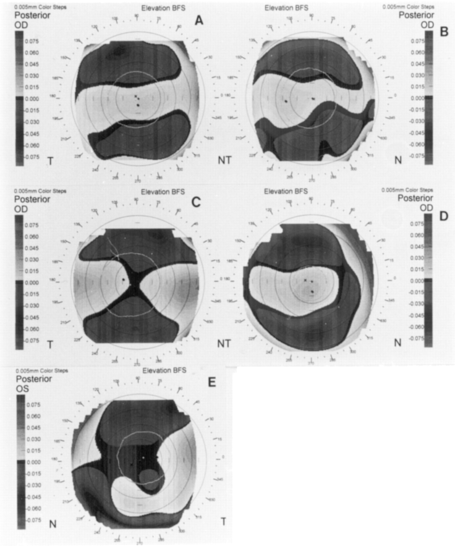
Elevation patterns of the posterior corneal surface: (A) regular ridge; (B) irregular ridge; (C) incomplete ridge; (D) island; (E) unclassified.
Because there was no existing method for classifying corneal pachymetry maps, the following classification system was devised. The warmest colour in the pachymetry map, identifying the thinnest area of the cornea, was used to designate one of four different patterns: round, oval, decentred round, and decentred oval. These patterns are depicted in Figure 2. The following objective criteria defined the categories used for this classification:

Pachymetric patterns in maps generated with the Orbscan corneal topography system: (A) oval; (B) round; (C) decentred round; (D) decentred oval.
Round: the ratio of the shortest to the longest diameter of the warmest colour zone was two thirds or greater, and the warmest colour was either entirely located at the centre of the map or more than half of the area of the warmest colour entered the central 3 mm diameter zone.
Oval: the ratio of the shortest to the longest diameter at the warmest colour zone was less than two thirds, and the warmest colour was either entirely located at the centre of map or more than half of area of the warmest colour entered the central 3 mm diameter zone.
Decentred round: the warmest colour was round, and more than half of its area was outside of the central 3 mm diameter zone or it was entirely located in the peripheral cornea.
Decentred oval: the warmest colour was oval, and more than half of its area was outside the central 3 mm diameter zone or it was entirely located in the peripheral cornea.
STATISTICS
Statistical analysis was performed by Mr William Feuer, a biostatistician in our department. The mean and standard deviation of the mean was used for various descriptive quantities. Difference in topographic measurements between eyes was calculated usingt test.
Results
A total of 94 eyes of 51 subjects (both eyes of 43 subjects and one eye of eight subjects) were investigated using the Orbscan corneal topography system. The fellow eye of eight subjects was excluded from this study was because of previous surgery (four eyes), contact lens wear (three eyes), and eyelid abnormality (one eye). The mean age of these subjects was 47.32 (SD14.06) years (range 23–78 years). The right eye of normal subjects (n=46) was used for the statistical analysis and the classification of topographic patterns.
The mean simulated keratometry (SimK) measurement was 44.24 (1.61)/43.31 (1.66) dioptre (D), which was not statistically different (p>0.2) from the mean SimK values in a study of normal corneal topography with the TMS-1 reported by Bogan et al.6 The mean keratometric astigmatism was 0.90 (0.41) D, which was also not significantly different from that reported by Bogan et al 6 (p>0.1) and another investigation of normal corneal topography with PAR Corneal Topography System reported by Naufal et al 5 (p >0.5). Mean astigmatism in the 3 mm, 5 mm, and 7 mm diameter zones was 1.22 (1.08) D, 0.94 (0.44) D, and 1.51 (0.91) D, respectively. Table 1 shows the astigmatism, irregularity, and mean keratometric power of the anterior and posterior corneal surfaces and the combination of the anterior and posterior corneal surfaces in the 3 mm, 5 mm, and 7 mm diameter zones. There was a decrease in the refractive power of the cornea and an increase in the irregularity from the centre to the periphery of the cornea in both the anterior and posterior corneal surfaces. No significant difference was noted in all of above factors between eyes.
Distribution of astigmatism, irregularity, and mean power in anterior and posterior corneal surface and combination of anterior and posterior corneal surface in 3 mm, 5 mm, and 7 mm diameters of 46 subjects
The most common anterior corneal elevation pattern was the island (71.74%), followed by incomplete ridge, regular ridge, irregular ridge, and unclassified patterns (Table 2). The island was the most commonly observed posterior corneal elevation pattern (32.61%), followed by regular ridge, incomplete ridge, and irregular ridge (Table2). No unclassified pattern appeared in posterior corneal elevation maps. The symmetric bow tie was the most commonly observed topographic pattern in the axial power maps of anterior corneal surface (39.13%), following by oval, asymmetric bow tie, round, and irregular (Table3).
Elevation pattern of the anterior and posterior corneal surfaces in the right eye of 46 normal subjects
Axial power pattern of anterior corneal surface in the right eye of 46 normal subjects
The thinnest site on the entire cornea was an average of 0.55 (0.03) mm thick and was located at an average of 0.90 (0.51) mm from the visual axis. This site was most commonly located in the inferotemporal quadrant (69.57%), followed by the superotemporal, inferonasal, and superonasal quadrants (Fig 3). Among the nine regions evaluated, the central cornea was found to be the thinnest (0.56 (0.03) mm). The superior cornea was the thickest (0.64 (0.03) mm), followed in order of decreasing thickness by the superonasal, inferonasal, inferior, superotemporal, nasal, inferotemporal and temporal zones (Fig 4). Pachymetry colour coded maps were classified into round, oval, decentred round, and decentred oval patterns. The majority of eyes had either oval (47.83%) or round (41.30%) patterns (Table 4). There was no statistical difference in corneal thickness of the thinnest site or the central and mid-peripheral measured sites between eyes.

Location of the thinnest site on the cornea measured by the Orbscan corneal topography system (n=46).

Average (SD) corneal thickness of 46 normal corneas centrally and at eight mid-peripheral locations
Pachymetrical pattern in the right eye of 46 normal subjects
Discussion
Clinicians have a number of methods for measuring corneal power and/or corneal shape including keratometry, keratoscopy, photokeratography, interferometry, computer assisted videokeratography, and rasterstereography.7-12 Before the introduction of the Orbscan corneal topography system, placido based computered assisted videokeratoscopy and rasterstereography were the only commercially available modalities to clinically evaluate corneal topography. The computered assisted videokeratoscope uses a collimating cone to reflect 25 or 30 rings off the corneal surface.6These reflected rings yield as many as 8000 data points for computer analysis. This technique detects curvature and refractive power of the anterior corneal surface. The PAR corneal topography system, uses stereorasterography to evaluate corneal surface topography.11 This instrument projects a grid of light onto a fluorescein dyed tear film covering the corneal surface. The fluorescent grid is then photographed at a defined angle, and the surface elevation topography is determined directly from distortions in the projected grid relative to a reference plane. The curvature and dioptric power of the corneal surface can also be calculated from these data.
The Orbscan corneal topography system is a new device to evaluate corneal topography by measuring the anterior and posterior corneal surface simultaneously.13 It provides both anterior and posterior corneal elevation maps as well as axial power data. This system yields more information about corneal refraction than placido and rasterographic systems because the refraction of posterior surface is also calculated. The shape of posterior surface may be important for detecting some corneal diseases—for example, posterior keratoconus, a condition that can only be diagnosed by evaluating the shape of the posterior corneal surface.
Corneal thickness can be evaluated by a number of methods including ultrasonic pachymetry,14-16 optical slit lamp pachymetry,16 specular microscopy,17 confocal microscopy,18 19 and partial coherence interferometry.20 Each of these methods has different clinical disadvantages. Large discrepancies in optical pachymetry results can be obtained by different observers or with different instruments. Ultrasonic measurement requires corneal contact and it is difficult to locate accurately the same points of measurement in serial examinations. This may result in falsely large variation in corneal thickness measurement. The Orbscan corneal topography system evaluates corneal thickness across the entire corneal surface and it is non-invasive. Using the Orbscan corneal topography system software, the standard location of central and periphery cornea can be designed according to clinical requirements. The pachymetry data in the present study are consistent with previous report by Herndon and associates.21 Yaylali and associates22reported that the relative accuracy and precision of the Orbscan system is similar to ultrasonic pachymetry, although they found that measurements of the corneal thickness with the Orbscan system were 23–28 μm greater than those obtained by ultrasonic pachymetry. Preoperative pachymetric measurements of central corneal thickness made with an ultrasonic pachymeter in the PERK study were 20 μm thinner than the measurements made with the Orbscan instrument in present study.23 Therefore, the pachymetric measurements by the Orbscan instrument cannot be used interchangeably with ultrasonic pachymetry measurements. One possible explanation for the observed differences in corneal thickness with these two methods is that the Orbscan system is a non-contact method whereas ultrasonic pachymetry requires corneal contact. Another possibility is that the Orbscan system may also measure the hydrated mucous gel covering the corneal surface that has been reported to be up to 40 μm thick.24 Despite these discrepancies, the Orbscan corneal topography system appears to offer clinical utility for assessing corneal shape and thickness.
In conclusion, the results of this study provide normal standards for elevation and curvature topography as well as corneal thickness using the Orbscan corneal topography system. These data will hopefully prove useful for future comparative studies of different corneal diseases.