Article Text

Abstract
AIM To explore the use of multifocal electroretinograms (MERG) in detecting early changes in age related macular degeneration (AMD).
METHOD 15 pre-AMD or early AMD eyes showing retinal drusen or irregular fundus pigmentation with window defects by fluorescein angiography (FA) and mildly decreased visual acuity were examined and compared with their asymptomatic fellow eyes. 20 age matched normal eyes were included as controls. MERG was recorded by a Veris system (version 3.0) using a 103 hexagon stimulus and 218 second total recording time per eye. The first order kernel was used to calculate amplitudes and latencies in three configurations: the nasal and the temporal areas, the superior and the inferior areas, and six concentric rings centred on the fovea.
RESULTS There were no significant differences in the amplitudes and the latencies between the different regions (nasal versus temporal and superior versus inferior) of the retina as well as between the different groups of eyes (normal, pre-AMD or early AMD, and the asymptomatic fellow eyes) in each region. Using the concentric configuration, the foveal amplitude of pre-AMD or early AMD eyes was significantly suppressed when compared with the age matched control group and their average latency was longer in the fovea than in outer rings and significantly prolonged when compared with the normal control group. Similar changes in amplitude and latency were also observed in the asymptomatic fellow eyes.
CONCLUSION Significant abnormality in the foveal amplitude and the foveal latency of MERG could be detected in pre-AMD or early AMD eyes as well as their asymptomatic contralateral eyes, suggesting MERG as a sensitive tool in detecting early foveal abnormalities in AMD.
- electroretinogram
- multifocal electroretinogram
- age related macular degeneration
Statistics from Altmetric.com
Normal ageing retinas exhibit a spectrum of changes such as decreased density of foveal photoreceptors and retinal pigment epithelial (RPE) cells while age related macular degeneration (AMD) retinas display similar losses, with or without subretinal neovascularisation, including atrophy of the pigment epithelium but with drusen as an early indicator.1-3 This loss of photoreceptors may affect the electrophysiological responses from the retina. Fish and Birch showed that the amplitudes of the central responses in focal electroretinograms (ERGs) of AMD eyes correlated with Snellen's acuity measurements while the latency was less predictive.4 Mayer et al showed that foveal flicker sensitivity was reduced in “exudative AMD risk eyes.”56 Hence, foveal electrophysiological measurements may be a useful tool in detecting early AMD changes.
Sutter and Tran7 introduced a multifocal ERG (MERG) system, which records the responses of multiple areas of the posterior retina in one sitting. It has been used in the detection of functional deficiency in diabetic retinopathy,89 occult macular dystrophy,10 myopia,11 macular hole,12 retinitis pigmentosa,13 and glaucoma.14 To explore the possible use of MERG in detecting early changes in AMD, we examined the MERGs from both eyes of patients who showed unilateral pre-AMD or early AMD changes.
Methods
In this study, early AMD changes were defined as mild visual impairment, age over 50, and macular drusen. All cases of early AMD eye were confirmed by two ophthalmologists (Li and Tso) independently. Almost all drusen were of the hard, single, dry type except patients 182 and 218 (Table 1). Pigmentary changes were hypopigmentation and they were light yellow drusen-like but not outstanding. These early AMD cases were selected from approximately 1500 local residents over the age of 40 in a United Nation Adult Vision Study (Hong Kong) in 1997. Preliminary screening showed 35 participants with unilateral macular drusen, macular irregular pigmentation, and decreased vision (below 1.0). Twenty five subjects then came back 1 year later. In the end, the resulting 15 subjects agreed to have MERG and fluorescein angiography (FA).
Clinical and MERG (configuration C) data of pre-AMD or early AMD eyes
MERGs were recorded from two groups of subjects (normal and patient groups) and divided into three groups of eyes: (1) 20 eyes of age matched normal volunteers; (2) 15 eyes with pre-AMD or early AMD; and (3) 15 asymptomatic, normal appearing fellow eyes of patients in group 2. The study was approved by the human ethics committee of the Chinese University of Hong Kong and adhered to the Declaration of Helsinki.
Each individual signed a consent form before the study. Normal volunteers with no known systemic or eye diseases were included in this study. AMD patients with unilateral early macular drusen or unilateral fundus irregular pigmentation with RPE changes and visual acuity 0.7 or above were examined. No difference in age was noted between the normal subjects and the patients (50.6 (SD 5.7) v52.1 (3.7); p=0.412) or refractive error (−0.25 (2.31) Dv −0.36 (1.59) D; p=0.874). Each subject received full ophthalmological examination and MERG recording. For the patient group, the first examination also included the Amsler grid test, FA, fundus photography, and medical/ocular history taking. They were also asked to have another MERG recording after 3 months. The MERG was recorded by a Veris 3.0 system (EDI, San Mateo, CA, USA) according to the method of Bearse et al.15 A 103 hexagon pattern stimulus and 32 segments for recording were used. The external gain was at 50 K, low cut off at 10 Hz, and high cut off at 300 Hz. A Burian-Allen bipolar contact electrode was used. A corrective lens was placed in front of the examinee, and the test distance was adjusted according to the power of the corrective lens. For data analysis, the 103 local responses were grouped into three different configurations: (A) the nasal and the temporal areas, (B) the superior and the inferior areas, and (C) six concentric rings (R1–R6) centred on the fovea (Fig 1). All P1 amplitudes and N1 latencies of the MERG responses in the three configurations were calculated and compared. ANOVA and Dunn's analysis were used for statistical purposes.
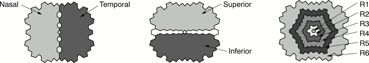
Configurations for the first kernel calculations of MERG. (Left) Nasal versus temporal retina. (Middle) Superior versus inferior retina. (Right) Concentric rings centring on the fovea.
Results
Of the 15 pre-AMD or early AMD eyes, nine of them showed a mild decrease in visual acuity. The mean is 0.84 ranging from 0.7 to 1.0 (Table 1). In contrast, the contralateral asymptomatic eyes showed normal visual acuity with a mean of 1.06, ranging form 0.8 to 1.5 (Table 2). Fundus examination showed a majority of the AMD group had hypopigmentation (eight out of 15), five eyes had one to three hard macular drusen, and two eyes had soft drusen (Table 1) while the fellow eyes were normal in appearance (Table 2). Twelve of 15 in the patient group received an FA examination of both eyes. Window defect or hyperfluorescence was seen in 11 pre-AMD or early AMD eyes (Table 1) and three fellow asymptomatic eyes (Table 2). With the Amsler grid test, central distortion, wavy lines, or blurred areas were noted in four pre-AMD or early AMD eyes (Table 1), with no abnormality noted in all asymptomatic fellow eyes (Table 2). Using configurations A and B, no significant differences were detected in the P1 amplitudes or the N1 latencies between the different regions (nasal versus temporal and superior versus inferior) of the retinas in the normal control eyes, the early AMD eyes, and the fellow eyes. There were also no significant differences in the P1 amplitudes and the N1 latencies with each region between normal, AMD, and the fellow eyes (Table 3). Contrary to configurations A and B, with configuration C, P1 amplitudes decreased and N1 latencies increased as the rings were further from the fovea in all three groups of eyes (Table 4). In the normal eyes, the foveal (R1) P1 amplitude was the highest among the six ring groups (p<0.001). However, there was no significant difference among the latencies of different rings. In the pre-AMD or early AMD eyes, the foveal (R1) P1 amplitude was also the highest among all six ring groups (p<0.001), and it was significantly reduced when compared with the normal (p=0.005). Foveal (R1) N1 latency was also the longest among the six rings (no significant difference) and was significantly longer than the normal (p<0.001). The fellow asymptomatic eyes showed values approximating those of the pre-AMD or early AMD eyes. The foveal (R1) P1 amplitude was also reduced compared to the normal (p=0.017), and the foveal N1 latency was also significantly longer than the normal (p<0.001). Figure 2 represents typical tracings of MERG responses in the six concentric ring configuration in the normal eyes, asymptomatic fellow eyes, and pre-AMD or early AMD eyes from two age and sex matched individuals showing the above mentioned features. Results of MERG at 3 months of follow up remained essentially the same (not shown) when compared with the initial examinations.
Clinical and MERG (configuration C) data of fellow asymptomatic eyes
Summary of P1 amplitudes and N1 latencies with configurations A and B
Summary of P1 amplitudes and N1 latencies with configuration C

Illustration of the MERG responses and their calculated P1 amplitudes and N1 latencies in (a) normal, (b) fellow, (c) and pre-AMD or early AMD eyes.
Discussion
In this study using the first kernel calculation of concentric ring configuration in MERG recordings, we noted a significant reduction in the P1 amplitude as well as a significant delay in the N1 latency of foveal responses from pre-AMD or early AMD eyes and their asymptomatic fellow eyes when compared with normal control eyes. These findings suggest that both foveal P1 amplitude and foveal N1 latency in MERG measurements may be sensitive means to detect early foveal AMD changes.
Configurations A and B were initially used because it was reported that in normal individuals the nasal retina showed a lower response than the temporal retina while there was no difference in the superior or inferior retina.16 However, we did not find any significant difference between these different regions of the retina or among the three groups of eyes.
Our observation that pre-AMD or early AMD eyes showed suppressed foveal P1 amplitude and prolonged foveal N1 latency is consistent with earlier reports noting foveal electrophysiological abnormality in AMD eyes.17 Contrary to other studies, we used eyes showing early or pre-AMD symptoms such as drusen and window defects in FA. Whether these eyes would ultimately suffer AMD remains to be determined but our study showed that foveal parameters of MERG are sensitive means to detect foveal changes.
Unlike the regular ERG, the exact origin of the first order kernel measurement is controversial although it is generally accepted that it arises from the cones.1819 The P1 amplitude of the central fovea is believed to be linked to the number and function of photoreceptor cells.1819 Our observation that the pre-AMD or early AMD eyes have a lowered P1 amplitude than their fellow eyes and the normal eyes is consistent with the hypothesis that the AMD eyes may have a reduced number and/or functional deficiency of photoreceptors. According to Kondo et al, the negative and positive deflections in the first order kernel of MERG probably do not correspond to conventional a and b-waves of ERG.20 Various investigators have suggested that MERG may include responses from the inner retina.2122 It is possible that the noted delay in the N1 latency reflects damages of the inner retinal layers as well as the outer retina. Although both the P1 amplitude and the N1 latency were affected, because of the smaller variations in latency measurements, the latter may be a more useful indicator of the changes.
Acknowledgments
We thank Michael Marmor, MD, of Stanford University, for commenting on the study. This study was supported in part by RGC grant CUHK405/95M, Hong Kong.