Article Text

Abstract
Aim: To report the authors' experience with the use of opaque intraocular lenses (IOL) in diplopia unresponsive to traditional management strategies, and to assess patients’ satisfaction and the visual function of patients following insertion of opaque IOL using the Visual Function Index (VF-14).
Settings: Royal Victoria Infirmary, Newcastle upon Tyne, UK.
Method: Data were obtained on all patients who underwent insertion of an opaque IOL at our institution between 2002 and 2006. Patients were interviewed by telephone. Any visual function impairment was assessed using the VF-14 questionnaire. Patients were also asked to score subjectively their overall satisfaction with the visual outcome after opaque IOL insertion.
Results: The authors studied 12 patients (n = 12) who had insertion of opaque IOL. All patients had constant and persistent diplopia unresponsive to other treatments acceptable to the patient. The median duration of diplopia was 5.5 years (interquartile range was 2.4–17.3 years). The postoperative VF-14 ranged from 75 to 100, and the mean VF-14 was 91 (95% CI 83–99). Three patients reported a maximum score of 100. Patient satisfaction ranged from 2 to 4, and the average was 3.4 out of 4.
Conclusions: Opaque IOL insertion is a valuable option in the management of intractable diplopia. The VF-14 revealed very little or no impairment in visual function following the procedure. All patients reported improvement in their visual function and were pleased with the final outcome.
Statistics from Altmetric.com
The onset of binocular misalignment in visually mature individuals causes diplopia (simultaneous perception of different images by the two foveas) or visual confusion (simultaneous perception of different images by retinal areas which normally correspond).1 These symptoms may be ameliorated or cured by the use of prismatic spectacle correction, injection of eye muscles with botulinum toxin,2 or by eye muscle surgery. A significant minority of patients, however, fail to respond to these strategies and have to occlude the vision in one eye to relieve their symptoms. Occlusion may be accomplished by patches, worn on the face or on spectacles; frosting or filters placed on the spectacle lens; or opaque contact lenses. Some patients with diplopia cannot tolerate these treatments or have persistent symptoms despite them. Opaque intraocular lens (IOL) insertion is a recently described option for occlusion.
To our knowledge, there have been only two case reports on the use of opaque IOLs in intractable diplopia, and we are unaware of any published data examining patients’ postoperative visual function.
We report our experience with the use of opaque IOLs in patients with diplopia unresponsive to other management strategies, and have investigated their visual function using the VF-14 questionnaire (a patient-reported measure of functional disability related to vision based on 14 everyday activities developed for use in patients with cataract).
METHODS
Data were obtained on all patients who underwent insertion of an opaque IOL at our institution between 2002 and 2006. Cases were identified from departmental electronic patient database (Mermaid) and operating theatre records.
Procedure
The crystalline lens of the eye with either worse visual acuity or greater restriction of ocular motility was removed. An opaque IOL (Morcher GmbH, model 81D diplopia lens, optic diameter 7 mm, overall diameter 13.50 mm, non-foldable) was inserted through a corneal wound. The wound was then sutured using 10/0 nylon. All patients had subconjunctival injection of antibiotics at the end of the operation and a topical steroid and antibiotic preparation was used for 4 weeks.
Data collection
Information was obtained from patients’ notes on age, gender, cause and duration of diplopia, relevant ophthalmic history, alternative occlusion measures attempted, visual acuity, evidence of sensory fusion and details of the surgery along with any complications.
VF-14
Patients were interviewed by telephone. The first two investigators (OHH and NW), who were not directly involved in the patients’ care, conducted the interviews. It was made clear to the patients at the beginning of the interview that the interviewer, although not completely independent, was not directly involved with their care in an attempt to minimise any bias. Any visual function impairment was assessed using the VF-14 questionnaire.3 Only activities that the patient considered relevant to their situation were scored. The patient was asked how much difficulty they had in performing each activity. There were five possible scores for each question: 0 (unable to do), 1 (great deal of difficulty), 2 (moderate difficulty), 3 (little difficulty), and 4 (no difficulty). A question was excluded if the patient did not perform the activity for reasons other than their vision. An average score was then generated from all the answered questions and multiplied by 25 to give a scale ranging from 0 (maximum disability) to 100 (no disability). At the end of the telephone interview, each patient was asked to score subjectively their overall satisfaction with the visual outcome after opaque IOL insertion. They were given a score scale from 0 (unhappy) to 4 (very satisfied).
RESULTS
Patient characteristics
Twelve patients (n = 12) were identified. The mean age was 43 (16.3) years; five were male, and seven were female.
Characteristics of diplopia
All patients had constant and persistent diplopia unresponsive to other treatments acceptable to the patient. The median duration of diplopia was 5.5 years (interquartile range (IQR) was 2.4–17.3 years). Four patients had diplopia for 4–10 years, and three patients had diplopia for more than 10 years. Eight patients developed diplopia following non-paralytic concomitant strabismus surgery. Four patients developed diplopia as a result of acquired paralytic incomitant strabismus. Two cases were traumatic, and the other two were secondary to an intra-cranial space-occupying lesion.
The Snellen best corrected visual acuity prior to insertion of IOL ranged from 6/5 to “Hand Movement” (the mode was 6/36) and in the fellow “normal” eye from 6/5 to 6/12 (mode was 6/6) (table 1).
Measures tried before opaque intraocular lens insertion
All patients had tried other occlusive methods before opaque-IOL insertion was contemplated. Blenderm/Bangerter filter was used in eight patients, and Fresnel prism was attempted in four patients. Occlusive contact lenses were tried in six patients with primary failure in two patients due to intolerance and persistence of diplopia. Initial improvement with occlusive contact lenses was reported in four patients over a period ranging between 1 and 8 months. Causes of discontinuation were reported as intolerance, irritation, headache, corneal epitheliopathy, and corneal ulcer. Rectus muscle chemodenervation with botulinum toxin to realign the eyes was undertaken in four patients and resulted in transient improvement. Diplopia corrective strabismus surgery was attempted in five patients, with unsuccessful outcome.
Two patients had early cataractous changes in the preoperative assessment, while the remaining 10 patients had a clear lens. The operation was performed under local anaesthetic in seven patients. Five patients requested general anaesthesia. Phacoemulsification was the technique used to remove the lens in 10 patients, and the other two patients had extra-capsular cataract extraction. Opaque IOL placement was in the capsular bag in 10 patients and in the sulcus in two patients.
The leading haptic of the opaque IOL broke at the haptic–optic junction during insertion in one patient. Successful secondary sulcus implantation was performed 6 months later.
All patients had an uneventful postoperative recovery. The postoperative visual acuity ranged from “hand movement” in three patients to “perception of light” in nine patients. Persistent postoperative diplopia was reported in one patient, which settled with topical pilocarpine 1%. Six patients have been discharged, and three are still been followed up with an annual ultrasound B-scan. Three patients are still under follow-up for unrelated ocular conditions in the operated eye including a previous episode of acute glaucoma, recurrent corneal ulcers, and band keratopathy.
Visual impairment and patients’ satisfaction
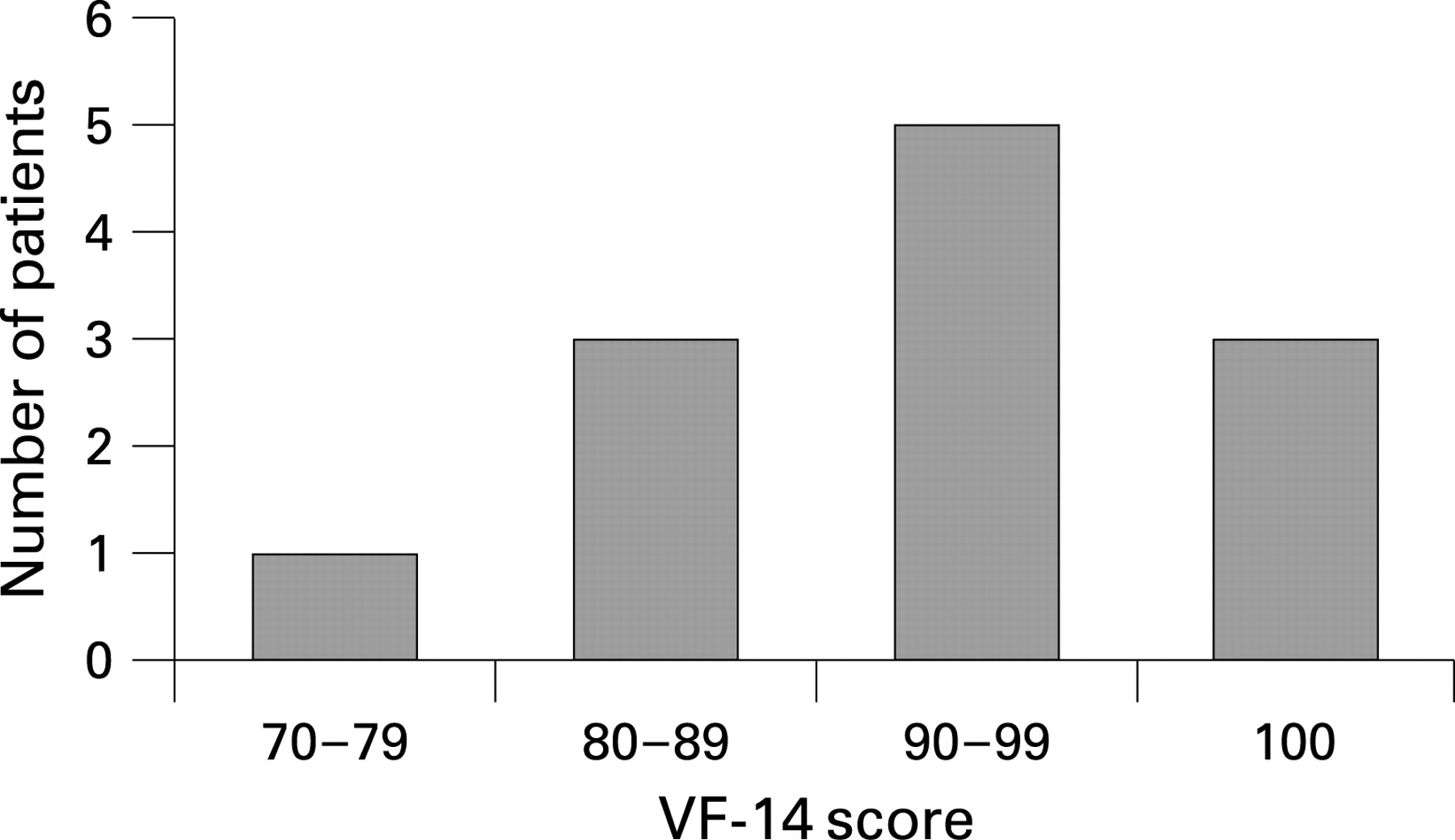
The average interval between the time of the operation and the date of the VF-14 assessment was 21 (12) months and ranged from 6 to 45 months. Figure 1 shows the VF-14 distribution following opaque IOL insertion. Table 2 summarises the individual patient response to VF-14 questions with their overall satisfaction. The reported postoperative VF-14 ranged from 75 to 100, and the mean VF-14 was 92 (95% CI 83–99). Only one patient reported a VF-14 of 75, and three patients reported a maximum score of 100.
Patient satisfaction ranged from 2 to 4, and the average was 3.4. Six patients scored 4 out 4, and only one patient scored 2 out of 4.
DISCUSSION
Binocular intractable diplopia is a rare but disabling condition. It is typically encountered following strabismus surgery or with paralytic strabismus.4 Management of intractable diplopia is complicated, as it is often resistant to standard treatment modalities. IOL insertion is a newer option, and to our knowledge this is the largest reported series looking at opaque IOLs in the literature.
There are limitations with all the above-mentioned treatment modalities for intractable diplopia, and it is clear that most of them are cosmetically unacceptable. Our patients achieved very little or short-lived relief with occlusive contact lenses, which goes in line with the reported literature. In one report on the use of opaque contact lenses, only 27% of patients with intractable diplopia found them a satisfactory form of treatment.7 These patients form a heterogeneous group who may have previously undergone multiple unsuccessful squint surgeries. Therefore, patients were reluctant to undergo further strabismus surgery. Botulinum toxin chemodenervation was attempted unsuccessfully in one-third of our patients due to the unpredictability and variability of the results.
Opaque IOL insertion, although more invasive than many of aforementioned treatments, has been used with some success by Sandy et al in their two case reports.5 Krieger et al have also recently reported a case were they successfully controlled a longstanding intractable diplopia with opaque IOL.6 The relative ease and availability of phacoemulsification with lens implantation coupled with a good safety profile means that this procedure is a valid and reasonable choice for intractable diplopia.
Preoperative counselling is crucial, and patients have to fully understand that the procedure is essentially irreversible and that monitoring or detecting posterior pole pathology in the operated eye is very difficult. Permanent uniocular occlusion may hinder the assessment of progression of visual field loss in patients with intracranial pathology. Insertion of an opaque IOL is contraindicated in patients who require monitoring of their posterior pole—for example for diabetic retinopathy.
One patient reported diplopia postoperatively despite having a poor recorded vision of “hand movements” in the operated eye, which resolved with pupil constriction. Three patients had poor preoperative vision of “counting fingers” or worse but were still symptomatic enough to agree to surgery and derived benefit. This raises interesting questions about the nature of diplopia symptoms and individual patient perception of diplopia and invites further study.
Our experience of a lens breakage requiring a delayed secondary implant highlights the importance of keeping a spare lens in case of unforeseen problems with the original implant.
The follow-up arrangements for patients after opaque IOL insertion varied in our series. Regular posterior pole ultrasound scans to identify any pathology is an option, but due to the relative infrequency of this occurrence, it may not be strictly necessary. Half of our patients were discharged from routine follow-up and were given clear instructions to seek medical help should they notice any symptoms pertaining to the operated eye. We do agree with Sandy et al that ultrasound scanning is also indicated in the early-postoperative period in patients at a higher risk of developing retinal detachment or if the postoperative course was stormy.
Newer designs are also improving the safety profile, and opaque IOL are now available in a foldable form which facilities smaller incisions. Phakic anterior chamber opaque IOL is a new option, and the relative ease of removal would render this procedure more easily reversible.
The VF-14 is a well-validated vision-related quality-of-life scale with high internal consistency,3 strong test–retest reliability8 and high responsiveness to change.9 10 The overall self-rating of the amount of difficulty patients have with their vision has also been shown to be more strongly correlated with the VF-14 than with measures of either visual acuity or generic health-related quality-of-life scales such as the SF-36.9 11
The VF-14 revealed very little or no impairment in visual function following the procedure. The mean VF-14 for our series was 91%, which was similar to the postcataract extraction score.12 All patients reported improvement in their visual function and were pleased with the final outcome. This is despite the fact that we are effectively “blinding” a healthy eye in many cases and this indicates how debilitating patients find intractable diplopia. A problem we would like to highlight, being a retrospective study, is the failure to record a preoperative VF-14 score. This would have been helpful to highlight any differences. Although the interviewers were not directly involved in the patients’ care, they were not from an independent body, so the possibility of some bias remains.
At first glance, it may seem counterintuitive to be “blinding” patients, and as such this is not a procedure that we would recommend lightly. However, reviewing our experience with this very select group of patients, we conclude that in the appropriate setting, this is a valuable option in management of patients with intractable diplopia. Prospective studies assessing their efficacy are now needed to further establish the role of opaque IOLs in this context.
Acknowledgments
The authors would like to thank M Dayan for performing the operation on patient 5.
REFERENCES
Footnotes
Competing interests: None.
Ethics approval: As this study was a service evaluation, formal ethics committee approval was not required.
Patient consent: Informed consent was obtained from all patients prior to surgery emphasising the risk of delaying or missing a diagnosis of primary posterior pole pathology.