Article Text

Abstract
Aims To comprehensively study the gross anatomy of human corneal innervation.
Methods Twenty-one specimens, including 12 normal human corneas from seven deceased patients, two eye-bank corneo-scleral buttons, two eye-bank corneo-scleral rims and five post-surgical specimens from three patients with keratoconus were studied. Corneal whole mounts were stained for cholinesterase enzyme using the Karnovsky & Roots direct colouring thiocholine modification of acetylcholinesterase (AchE) technique.
Results Approximately 44 thick nerve bundles were found to enter the human cornea in a relatively equal distribution round the limbus and move randomly towards the central cornea. At the mid-peripheral zone, anterior stromal nerves showed a characteristic budding and branching pattern. After passing through Bowman's zone they were noted to terminate into bulb-like thickenings from which multiple sub-basal nerves arose. The perforation sites were predominantly located in the mid-peripheral cornea. The orientation of sub-basal nerves was mainly vertical at their origin from the perforation sites. Nerves from all directions converged towards the infero-central cornea to form a characteristic clockwise whorl pattern.
Conclusions This study provides a comprehensive account of the architecture and distribution of nerves in the human cornea. It reconciles some of the existing information obtained from other modalities of investigation and identifies some novel features that provide a more complete picture of corneal innervation.
- Cornea
- nerves
- histochemistry
- cholinesterase
Statistics from Altmetric.com
Introduction
The human cornea is one of the most richly innervated structures in the body and is densely supplied by sensory and autonomic nerve fibres.1 The sensory nerves, which constitute the majority of corneal nerves, are mainly derived from the ophthalmic division of the trigeminal nerve.2–8 They have a variety of sensory and efferent functions. Mechanical, thermal and chemical stimulation of the corneal nerves produce predominantly a sensation of pain in humans.9 The autonomic nerve fibres consist of sympathetic fibres that are derived from the superior cervical ganglion10 and parasympathetic fibres that originate from the ciliary ganglion.11–13 Corneal sensation is essential for maintaining the integrity of the ocular surface.
Different techniques have been used to demonstrate corneal nerve patterns in different mammalian corneas.14–17 In 1951, Zander and Weddell published the earliest detailed observations on the innervation of the mammalian cornea.18 However, it provided relatively limited knowledge on human corneal nerves.
The effect of cutting and laser ablation of the cornea in various refractive surgery procedures has drawn much attention to the corneal innervation in recent years. Some authors suggest that the corneal nerves enter the cornea predominantly at the 3 and 9 o'clock positions; thus creation of a nasally hinged flap for laser-assisted in situ keratomileusis (LASIK) surgery preserves half of the nerves, resulting in less corneal nerve damage and dry-eye-related postoperative complications.19 20 Rapid recovery of corneal sensation following LASIK is also attributed to the preservation of nerves in the nasal hinge.21 However, Kumano et al have shown the opposite results, where corneal sensation was less in eyes undergoing LASIK with a nasally hinged flap.22 A recent study (using IntraLase femtosecond laser) revealed insignificant differences in the corneal sensitivity and incidence of postoperative dry eye complications between superior and temporal hinged flaps.23 The notion that positioning of the flap can limit corneal nerve damage and its consequences probably reflects our limited understanding of corneal innervation.
Muller et al reported that nerve fibre bundles in the sub-basal plexus across the central and mid-peripheral cornea run first in the 3–9 o'clock hours direction, then after bifurcation, travel in the 6–12 o'clock hours direction and after a second bifurcation again in the 3–9 o'clock hours direction.17 However, more recent human studies with in vivo confocal microscopy (IVCM) suggest an alternative model of central sub-basal corneal innervation, where leashes of nerve fibres extend across the corneal apex preferentially in the 6–12 o'clock direction and other leashes approach the apex in the 5–11, 1–7, 3–9, 2–8 and 4–10 o'clock directions.1 24–26 Due to technical reasons, IVCM images are limited mostly to the corneal apex and cover a small area of the central cornea. Consequently, the orientation of sub-basal nerves in peripheral regions of the human cornea remains incompletely understood. In addition, there are few data on the topographical distribution of stromal innervation in human cornea.
We therefore undertook to verify comprehensively the normal anatomy of the central and peripheral human corneal nerves using a whole mount cholinesterase method, which has been proven to be excellent for quantitative and qualitative analysis of corneal nerves in different species, for example rats, rabbits and dogs.15 27–29 There is limited reference to this method on human samples (to study graft re-innervation).30
Materials and methods
Materials
Twelve normal human corneas from seven deceased patients (four women, three men) were examined. Mean age was 76.6 years. These eyes were obtained at different time intervals after death. Two eyes were obtained within 24 h post mortem, four eyes (two pairs) between 24 and 48 h post mortem and six eyes (three pairs) between the second and third post mortem days.
Two eye-bank corneo-scleral buttons maintained in organ culture. One was processed 5 weeks post mortem and the other 13 weeks post mortem.
Two eye-bank corneo-scleral rims maintained in organ culture, 3 weeks and 19 weeks post mortem.
Five post-surgical corneal specimens were obtained from three patients (aged 18, 28 and 38 years) with advanced keratoconus who underwent deep anterior lamellar keratoplasty (DALK) using the big-bubble technique. The superficial (approximately 50–70% thickness) and deep lamellae were studied. It has been shown that while the central corneal nerves are altered, the mid-peripheral and peripheral corneal nerves remain unaffected in keratoconus.31
The orientation of the enucleated eyes was marked by leaving part of the superior rectus muscle intact in order to mark the 12 o'clock position. Immediately after enucleation, the eyes were placed in phosphate buffered saline at 4°C (Dulbecco's PBS; Sigma-Aldrich, Poole, UK). The corneas were isolated by a circumferential cut along the limbal margin leaving a 2–3 mm scleral rim surrounding the cornea. The scleral rim was fashioned such that the 12 o'clock position could be identified in the corneal scleral disc. The orientation for eye-bank eyes in organ culture could not be ascertained. Corneas were fixed in 4% formaldehyde (pH 7) for 4 h and then rinsed overnight in PBS. All corneas were stained non-specifically with Karnovsky & Roots direct colouring thiocholine modification of acetylcholinesterase (AchE) technique.32 Briefly, the staining solution was freshly prepared as shown in table 1.
Acetylcholinesterase-staining solution preparation
All specimen were incubated in the stock solution for 24 h at 37°C. The incubation solution was used without enzyme inhibitor to permit non-specific acetylcholinesterase (NsAchE) staining. The staining reaction was arrested by rinsing in three changes of PBS with a 15–20 min interval between rinses. The reaction product was intensified with a dilute ammonium sulphide (two drops of NH2S in 10 ml de-ionised H2O) rinse for 8 min, followed by two consecutive 5 min deionised water rinses. Specimens were secured between two glass slides and immersed in 80% alcohol for 1 day, followed by five changes of absolute alcohol over the next 5 days. Clearing, to remove alcohol, was achieved with seven changes of xylene over the next 7 days. The specimens were mounted between two slides and weighted for 24 h to allow the mount to set. A normal rectal biopsy specimen was used as a positive control.
All photomicrographs were taken using an Olympus BX40 microscope and a DP12 Olympus camera (Olympus, Tokyo, Japan). Adobe PhotoShop CS (Adobe Systems Inc., San Jose, California, USA) software was used to construct a montage of corneal nerve images. For determining topographical differences in innervation, each corneal specimen was examined in four pie-shaped quadrants: superior (10.30–1.30 o'clock), inferior (4.30–7.30 o'clock), nasal right eye and temporal left eye (1.30–4.30 o'clock), and temporal right eye and nasal left eye (7.30–10.30 o'clock). Limbal nerves entering each quadrant were counted.
Results
Origin and architecture of sub-basal nerve plexus
AchE-positive sub-basal nerves were demonstrated only in corneas harvested within the first two post mortem days and in the post-surgical specimens. They appeared as linear structures running in the superficial layer of the cornea with frequent Y-shaped bifurcations and re-unions or unions with other branches and contained densely stained fine granular structures (figure 1A). These nerves originated from the sub-Bowman's nerves that were located in the most anterior part of the corneal stroma. Theses nerves appeared to penetrate the Bowman's layer perpendicularly giving rise to multiple sub-basal nerves (figures 1B and 1C). Interestingly, some of the thicker sub-Bowman's nerves divided into two (figure 1D) or more branches just before perforating Bowman's layer giving a characteristic ‘budding and branching pattern’ (figures 1E). This branching pattern was more evident in the mid-peripheral cornea. A novel finding was that the perforation sites were mainly located in the mid-peripheral cornea and characterised by densely stained disc or bulb-like thickenings from which the sub-basal nerves arose (figure 1F). Relatively fewer perforation sites were found in the central and peripheral cornea. Perforation site counts were done on two whole montages. In one corneal button, there were about 25 perforation sites in the central cornea and 160 in the mid-peripheral cornea. Another button showed about 30 perforation sites in the central cornea and 125 in the mid-peripheral cornea (figure 2).
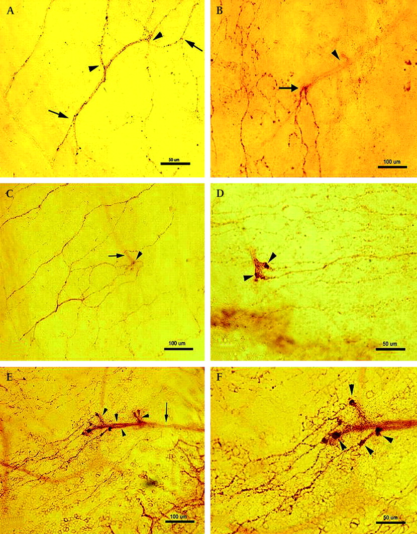
Photomicrographs of whole human corneal mount stained by the acetylcholinesterase technique. (A) sub-basal nerve plexus with characteristic branching (arrows) and union/re-union (arrowheads). The nerves contain densely stained fine granular material. (B) sub-basal epithelial leashes of nerves in a human cornea. The arrow shows the point at which a sub-Bowman's nerve penetrates Bowman's zone giving rise to multiple sub-basal nerves. The sub-Bowman's nerve is out of focus in this microscopic image (arrowhead). (C) A thicker sub-Bowman's nerve (arrow), which reaches the epithelium at the site of perforation (arrowhead) giving rise to multiple thinner sub-basal nerves. (D) A sub-Bowman's nerve bifurcates and penetrates to emerge anterior to Bowman's zone terminating in discoid or bulbous thickenings (arrowheads) which give rise to sub-basal nerves. (E) A single sub-Bowman's nerve (arrow) gives multiple branches (arrowheads) just before perforating the Bowman's zone ‘budding and branching pattern’. (F) A higher magnification of the same nerve in figure (E) showing characteristic bulb like thickenings at the perforation site (arrows) from which sub-basal nerves arise. Scale bars, 50 μm (A, D, F) and 100 μm (B, C, E).

Photomicrograph of whole human corneal mount stained by the acetylcholinesterase technique showing the perforation sites (marked with black dots), which are predominantly distributed in the mid-peripheral cornea. There are about 30 perforation sites in the central cornea (area within the circle) and 125 in the mid-peripheral zone (outside the circle) (montage of 37 images).
The directional orientation of the sub-basal nerves at their origin at the perforation sites was mainly vertical, that is running downward (superior to inferior) for nerves arising from perforation sites in the superior and central cornea and upward (inferior to superior) for those arising in the inferior cornea. The nerves also ran parallel to each other. Interestingly, the orientation of the sub-basal nerves arising from nasally and temporally located perforation sites, were also vertical, running downwards (superior to inferior) and obliquely towards the central cornea. Nerves from all quadrants converged towards the central cornea and formed a whorl pattern inferior to corneal apex (figure 3A). Moreover, the rotation of the sub-basal nerves in the region of the whorl was clockwise.
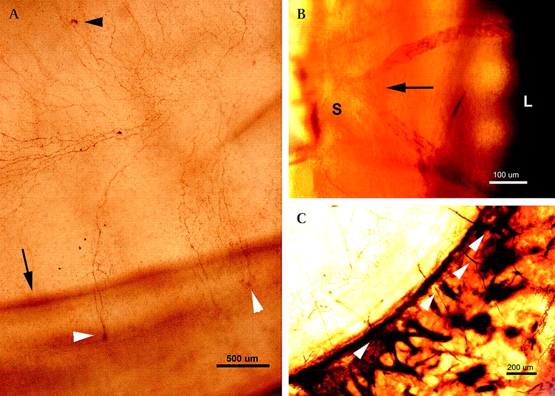
Photomicrographs of whole human corneal mount stained by the acetylcholinesterase technique. (A) A clockwise whorl pattern of the sub-basal nerves in the infero-central corneal region. Perforation sites of the sub-basal nerves in the inferior (white arrowheads) and central cornea (black arrowhead) were demonstrated. A dark brown line (arrow) represents a corneal fold due to the effect of flattening of the cornea during processing. (B) A pre-corneal nerve bifurcates (arrow) in the sclera (S) prior to its entry to the corneal limbus (L). (C) Nerves enter the corneal limbus as a direct continuation of nerves in the suprachoroidal space (white arrowheads). Scale bars, 500 μm (A), 100 μm (B), 200 μm (C).
Limbal and stromal nerves
AchE-positive stromal nerves were demonstrated in all corneal specimens. Nerves were found to enter the corneal limbus at different levels predominantly in the mid and deep stroma. Most of them were noted to be a continuation of the nerves in the suprachoroidal space (figures 3B & 3C).
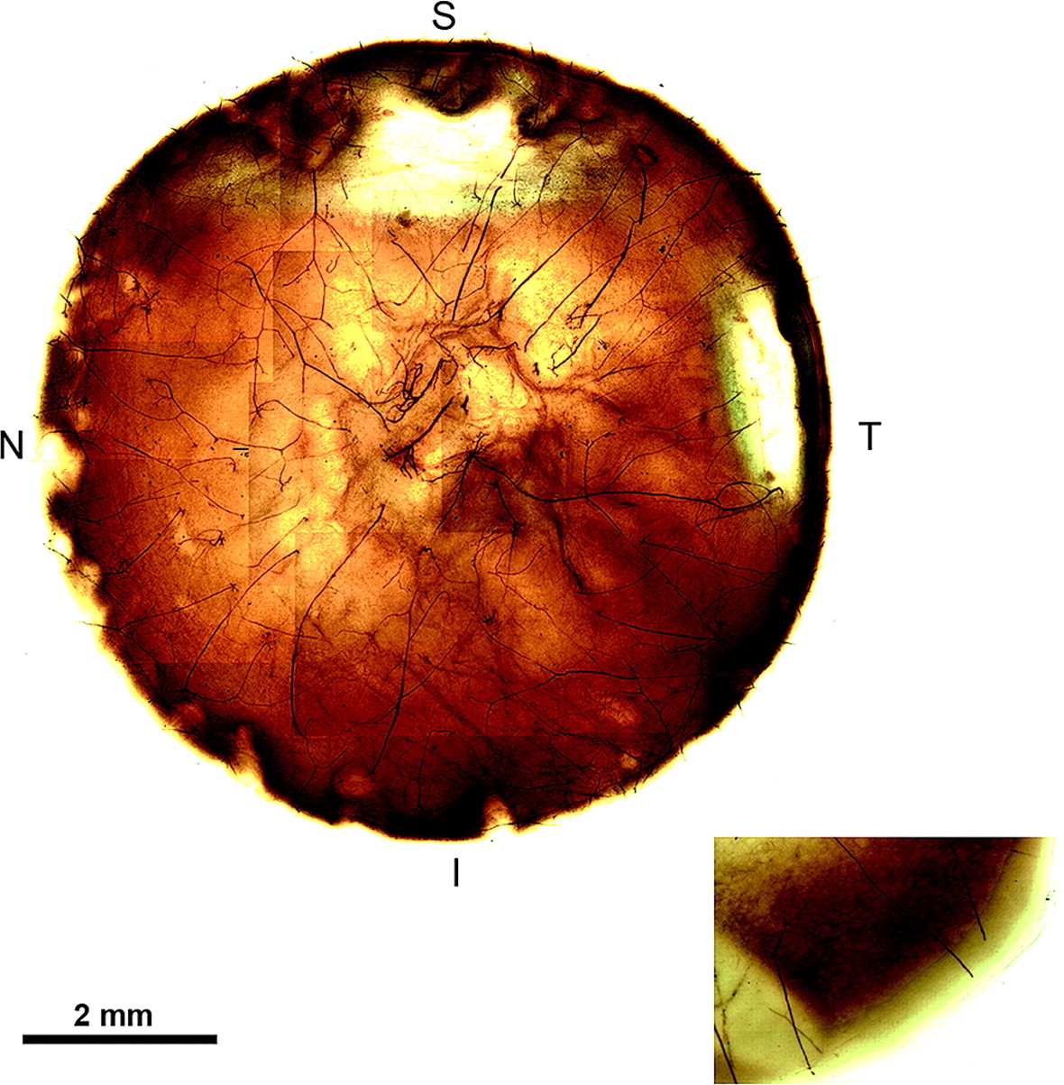
The numbers of limbal nerves entering each quadrant were counted and the average was as follows: superior (11.00), medial (9.43), inferior (11.43) and lateral (11.86) (figure 4). Overall, corneas were found to be innervated by an average of 43.72 AchE-positive thick nerve bundles, which were distributed evenly round the limbus. To obtain a holistic view of corneal nerve distribution, mapping of corneal nerves in the whole anterior lamella of post-surgical specimens was achieved by a montage of 37 images, and clearly showed that nerves were coming from all corneal quadrants (figure 5). Moreover, large nerve trunks were also shown in the posterior lamella, which represent approximately the posterior one-third of the corneal stroma (figure 5 inset).

A box plot showing the distribution of corneal nerves around the human limbus in four corneal regions (superior, medial, inferior and lateral). Means are indicated by solid circles.
Photomicrographs of whole human corneal mount of the left eye stained by the acetylcholinesterase technique A montage of 37 images giving a holistic view of the distribution of corneal stromal nerves in the anterior lamella of a deep anterior lamellar keratoplasty (DALK) corneal specimen. The posterior lamella also showed a similar pattern (inset). Scale bar, 2 mm (same specimen as figure 2). S, superior, I, inferior, N, nasal, T, temporal.
Nerves from eye-bank specimens retained enzymatic activity of cholinesterase even at 19 weeks post mortem and stromal nerves were visible peripherally and centrally.
Discussion
In this study, the direct colouring thiocholine modification of AchE technique was used to demonstrate human corneal nerves in whole-mount corneas. This technique allowed excellent in vitro three-dimensional visualisation of the distribution and spatial arrangement of the nerve bundles.
Over the past few years, several studies have been conducted to investigate the architecture and quantitative features of the human sub-basal nerve plexus.17 26 31 33–45 However, the anatomical relationship between sub-basal nerves and anterior stromal nerves in terms of the location and distribution of perforation sites and the behaviour of the nerves around these sites have not been elucidated and are novel to this study. To the best of our knowledge, this is the first demonstration (using the direct colouring cholinesterase method) of the overall and relative distribution of the perforation sites in central and peripheral human cornea wherein these sites are predominantly located in the mid-peripheral cornea. This correlates with a recent in vivo confocal study observation of probable sites of perforation of nerves through Bowman's layer, which appeared as an abrupt termination of the sub-basal nerves into bright, irregularly shaped areas 20 to 40 μm in diameter mainly observed in mid-peripheral cornea.31 Our study shows that these bright irregular structures commented on previously most probably represent the disc- or bulb-like thickenings observed at the sites of perforation. It also makes the definitive link between the sub-Bowman's nerves, their perforation to the sub-basal plane and termination in bulbous structures from where the leash of sub-basal nerves arise. The technical properties of confocal optics (small field of view and limited axial resolution) may not have allowed a complete understanding of the bright structures observed.
Schimmelpfennig,46 using a gold chloride impregnation technique, reported five to eight dichotomously branching sub-Bowman's nerves in central human cornea that gave off numerous sub-basal nerves and described this appearance as ‘leash’. However, the author did not comment on the directional orientation of the nerve fibres.
A recent electron microscopy study reported that perforation sites are mainly located in the central cornea in humans and suggested that this innervation, together with a conjunctival supply, contributes to human epithelial innervation.47 However, we were able to demonstrate fewer perforation sites in the central and peripheral cornea compared with the mid-peripheral zone. The bulbous structures found in close proximity to the perforation sites appear novel and densely stained, suggesting a high level of esterases in these structures. As they were shown in all corneal specimens, they probably represent normal anatomical structures rather than artefacts. Similar structures were also reported by Guthoff et al48 using fluorescent laser scanning confocal microscopy to detect the conversion of calcein acetoxymethylester into fluorescing calcein by esterases. They described them as 5–10 μm structures located just above the Bowman's layer. However, Guthoff et al48 had used corneas obtained by keratoplasty from patients with Fuchs endothelial dystrophy, and they suggested that these thickenings could either be related to Fuchs dystrophy or might resemble the bulbous thickenings in the rabbit cornea briefly mentioned by Elder in 1961.49 The fact that these structures have not been described in previous light and electron microscopy studies could be due to limitations of the techniques used, specimen size and the location of sampled area (most being limited to central cornea).1 17 46 47 50
Another interesting observation from our study was the budding and branching pattern of the sub-Bowman's nerves, which gave rise to two or more branches just before penetrating the Bowman's membrane. This corresponded with the perforation sites in the mid-peripheral zone. Such branching would allow for increase in the density of innervation of the more superficial sub-basal plexus leading to enhanced corneal sensitivity. Furthermore, the abundance of sub-Bowman's nerves and their branches in the mid peripheral region could explain the findings by Auran et al51 using IVCM. They described a ‘sub-epithelial plexus’ referring to nerves in the most anterior part of the stroma, which were sparse and patchy in distribution but most prominent at the mid-peripheral cornea. They also stated that this ‘sub-epithelial plexus’ may not be present in the central cornea.
We also noted that the orientation of the sub-basal nerve fibres, as they emerged from the perforation sites, was mainly vertical, that is running inferiorly and superiorly, rather than radial. However, as the sub-basal nerves from all quadrants converged towards the cornea apex they assumed a curvilinear orientation with a clockwise whorl-like disposition. A previous IVCM study has elucidated this whorl pattern31 in the central cornea, but due to the limitations of the technique their examination was confined to approximately the central 5×5 mm only and the findings cannot be extrapolated to the entire cornea. Our study (figure 3A) is the first histological demonstration of the whorl pattern of the central sub-basal nerve architecture. Interestingly epithelial whorl patterns, predominantly clockwise in orientation, have been reported before and may be related to the orientation of the sub-basal nerve plexus.52–54
We found this technique to be useful in visualising nerves in the suprachoroidal space in close vicinity to the human corneal limbus. This was helpful to demonstrate the origin of limbal nerve trunks, which were noted to be a direct continuation of the nerves running in the suprachoroidal space, in addition to those arising from the peri-limbal plexus. This pattern in humans is somewhat different from that of other species, where large corneal nerves were shown to originate from a complex peri-corneal nerve plexus.55
Staining of two posterior lamellae of DALK specimens revealed thick nerve bundles at the periphery. These specimens represent approximately 150 μm of the posterior corneal stroma suggesting that these nerves possibly enter the cornea at levels deeper than the mid-third of stroma. Further studies to determine the exact depth of entry of nerves are underway.
Using techniques other than cholinesterase staining, Zander and Weddell reported that about 70–90 nerve bundles enter the human cornea18 but they did not comment on topographical distribution. Quantitative analysis of corneal nerve distribution has been studied in rats and dogs27 28 but not in humans. In our study, we noted that innervation is largely by approximately 44 AchE-positive thick nerve bundles, which were distributed relatively evenly round the limbus. We did not see a preferential concentration of nerve bundles in the 3 and 9 o'clock meridian. Using the same technique, Lasys et al reported that dog's cornea is innervated by 12–15 thick nerve bundles, which are distributed equally round the limbus.28 As the stromal nerves were shown to be distributed evenly in all quadrants, LASIK flap creation will probably induce the same damage to the stromal nerves and their branches, as they approach the perforation sites, regardless of the position of the hinge. As described above, clinical studies19–23 have had contrasting findings in terms of flap position and corneal sensation. Our findings would push the evidence towards there being no difference on corneal sensation with respect to flap position.
AchE-positive limbal and stromal nerves were detected 3 months after death. This is interesting as it is well known that this technique is based on the enzymatic activity of acetylcholinesterase, which starts to decline very soon after death.56 In eye-bank eyes, the suitable storage medium and temperature may allow preservation of the enzyme.57 Sub-basal nerves could not be detected after 48 h post mortem, which may be due to a combination of nerve fibre degeneration and enzyme degradation sufficient to prevent visualisation of these fine nerves.
Our study provides a comprehensive account of the architecture and distribution of nerves in the human cornea. It reconciles some of the existing information obtained from other modalities of investigation and identifies some novel features that provide a more complete picture of corneal innervation and its clinical implications in humans. The description of the bulbous termination of sub-Bowman's nerves at the perforation sites and the origin of leashes of sub-basal nerves from these bulbous terminations is a uniquely novel finding of this study.
Acknowledgments
We thank Mr VS Maharajan (Cornea Consultant, Department of Ophthalmology, Queens Medical Centre, Nottingham, UK), and Janet Palmer and Katherine Fowkes Burchell (Department of Neuropathology, Queens Medical Centre, Nottingham, UK). The corresponding author (HSD) had full access to all the data in the study and takes responsibility for the integrity of the data and the accuracy of the data analysis.
References
Footnotes
Competing interests None of the authors have any proprietary/financial interest to disclose.
Ethics approval This study was conducted with the approval of the Nottingham Research Ethics committee. 07/H0403/140. The study adhered to the tenets of the declaration of Helsinki. The study was carried out in premises licensed under the Human Tissues Act, UK (2004).
Provenance and peer review Not commissioned; not externally peer reviewed.