Article Text

Abstract
Aims To describe baseline characteristics and visual outcome for eyes treated with ranibizumab for diabetic macular oedema (DMO) from a multicentre database.
Methods Structured clinical data were anonymised and extracted from an electronic medical record from 19 participating UK centres: age at first injection, ETDRS visual acuity (VA), number of injections, ETDRS diabetic retinopathy (DR) and maculopathy grade at baseline and visits. The main outcomes were change in mean VA from baseline, number of injections and clinic visits and characteristics affecting VA change and DR grade.
Results Data from 12 989 clinic visits was collated from baseline and follow-up for 3103 eyes. Mean age at first treatment was 66 years. Mean VA (letters) for eyes followed at least 2 years was 51.1 (SD=19.3) at baseline, 54.2 (SD: 18.6) and 52.5 (SD: 19.4) at 1 and 2 years, respectively. Mean visual gain was five letters. The proportion of eyes with VA of 72 letters or better was 25% (baseline) and 33% (1 year) for treatment naïve eyes. Eyes followed for at least 6 months received a mean of 3.3 injections over a mean of 6.9 outpatient visits in 1 year.
Conclusions In a large cohort of eyes with DMO treated with ranibizumab injections in the UK, 33% of patients achieved better than or equal to 6/12 in the treated eye at 12 months compared with 25% at baseline. The mean visual gain was five letters. Eyes with excellent VA at baseline maintain good vision at 18 months.
- Retina
- Macula
- Treatment Medical
- Treatment Surgery
- Public health
Statistics from Altmetric.com
Introduction
Intravitreal injections of ranibizumab are an established therapy to treat a sight-threatening complication of diabetic retinopathy (DR), diabetic macular oedema (DMO). In the UK, the second highest annual National Health Service (NHS) expenditure for any single drug is for ranibizumab (£244 million).1 Clinical practice was initially informed by pivotal clinical trials,2–7 which demonstrated that ranibizumab prevents central vision loss and improves mean visual acuity (VA), when given at monthly or according to pro re nata intervals in eyes with DMO.
In the UK, the National Institute for Health and Care Excellence approved the use of ranibizumab for DMO in February 2013, leading to universal availability of this drug in the NHS, but only if the central retinal thickness was 400 microns or more. Delivering the therapy with a recommendation of monthly assessment and then retreatment for ‘active’ disease makes substantial demands on healthcare services, funding authorities and patients. A recent review of the use of ranibizumab for any indication shows a more than twofold geographic variation in usage across England.8 The intervention is costly and intensive, so it is important to understand what visual outcomes are achieved and how clinical trial results ‘translate’ into clinical practice in the ‘real world’.
Clinical trials are limited by entry criteria, for example, excluding patients with uncontrolled medical conditions or severe DR, and have a limited number of trial subjects. In particular for DMO, the haemoglobin A1c and blood pressure in pivotal trials can be substantially different from ‘real-world’ patients,9 ,10 although a recent study suggests this may not affect outcomes of treatment.11 Electronic medical record (EMR) systems, if well-designed, offer more complete, prospective, real-time data collection as a by-product of routine care. They can be designed to mandate capture of a defined minimum dataset and allow the collection of more enriched datasets. The NHS Connecting for Health's ‘Do Once and Share’ programme has defined EMR datasets in ophthalmology including: diabetic eye disease, the cataract national dataset and the glaucoma dataset.12
This study aims to report the visual outcome and define benchmark standards of care for patients treated with ranibizumab for DMO at a large number of the UK centres.
Methods
Ethical approval
The lead clinician and Caldicott Guardian at each centre gave written approval for extraction of anonymised data. The study protocol was approved by the head of research governance at the lead clinical centre. This study was conducted in accordance with the Declaration of Helsinki and the UK Data Protection Act.
Data collection
Anonymised data were remotely extracted from 19 centres using the same EMR system (Medisoft Ophthalmology, Medisoft, Leeds, UK) in November 2014. Each site is the only NHS provider of DMO care to their local population and very few patients switch between providers or access care privately. Data were extracted through the EMR compulsory DR structured assessment module (see online supplementary file for detailed explanation). Demographic data were extracted from the hospital's patient administration system to the EMR.
supplementary file
All patients had data extracted from the time of their first DR structured assessment entry onto the EMR, including the data from the time of their first injection of ranibizumab up to the date of their last clinical entry before the data extraction on 26 November 2014.
Missing data
The EMR data extraction does not record values on visits where the EMR was not used, so no missing value substitutions were performed. The only exception to this rule was baseline VA. Some treatment centres operate ‘2 stop’ or ‘injection only’ clinics, where treatment is given without measuring vision. Six centres have over 10% of injections without recording a VA measurement on the same day. For these services, the baseline VA was taken from the prior assessment visit if within 4 weeks of the injection date. This was therefore not missing data per se but reflects variation in treatment delivery. Therefore, data on number of visits represent a normalised value to allow standardised comparison between centres, rather than the precise number of attendances by the patient. Analyses of all patients initiated into the study were compared with the cohort of patients that completed follow-up.
Analysis
The primary analysis was restricted to treatment-naïve eyes undergoing ranibizumab therapy as the only treatment for DMO during the follow-up period. Secondary analyses of eyes with other treatments prior to undergoing intravitreal ranibizumab therapy, or who were treated with combination therapy, were also undertaken. Eyes that had cataract surgery within 3 months of their first ranibizumab injection or during the period of follow-up were excluded.
Eyes were assigned to two groups according to their treatment history and were analysed separately. Group 1 eyes were treatment naïve at baseline for any treatment for DMO (including intravitreal injections of any drug, macular laser treatments and vitrectomy), but could have had previous peripheral scatter retinal laser at the time of their first injection of ranibizumab and were managed solely with ranibizumab during the course of follow-up until the end of the data extraction period. DMO group 2 patients had received other treatments for diabetic macular disease prior to their first injection of ranibizumab.
Results
Participants
Data were extracted on 123 968 eyes of 61 984 patients with DR. There were 33 967 male patients and 28 002 female patients (in 15 cases, gender was not recorded).
The 19 UK hospitals treated a total of 3103 eyes from 2416 patients, who received 15 537 ranibizumab injections for centre involving diabetic macular oedema (CIDMO) during 12 989 clinic visits. No patients in this dataset received ranibizumab for non-CIDMO. Of these, 28% (n=687) patients received bilateral treatments.
The 19 sites entered their first DMO ranibizumab treatment episodes into the EMR systems during the following years: 2008 (n=4 sites), 2009 (n=1), 2010 (n=5), 2011 (n=5), 2012 (n=3), 2014 (n=1). The first recorded ranibizumab injection for DMO was dated 10 June 2008.
The mean age (at the time of the first DMO injection) was 66 years (SD 13 years). The female to male ratio was 1.45:1 (1430/986 patients). There were 268 (16% of the patients with a known diabetic status) patients with type 1 diabetes mellitus. There were 1380 patients (84%) recorded with type 2 diabetes mellitus. The rate of endophthalmitis was 0.015%.
Treatment history
There were 1584 treatment-naïve eyes in group 1. Group 2 had 1471 eyes (1112 macular laser treatments, 77 vitrectomies, 332 bevacizumab, 212 with triamcinolone). Of the patients in group 2 who had received previous treatment, the median (IQR) time between the first ranibizumab injection and the previous diabetic macular treatment was 0.6 (0.2, 1.2) years.
Visual acuity
About 34.1% of best corrected visual acuity tests were converted by the EMR software at source from Snellen to letter score equivalent. The rest were recorded as ETDRS letters. Baseline VA is shown in online supplementary figure S1 for all treatment groups. There was a mean gain of five letters at 12 months when both groups were analysed.
Treatment-naïve eyes
The proportion of eyes with VA of 20/40 or better (72 or more letters) in the better-seeing eye was 25% at baseline, 33% at year 1, 24% at year 2. A more complete understanding of the change in vision is obtained by plots of VA at different time points (figure 1) demonstrating mean, and SD of the data stratified by starting VA.
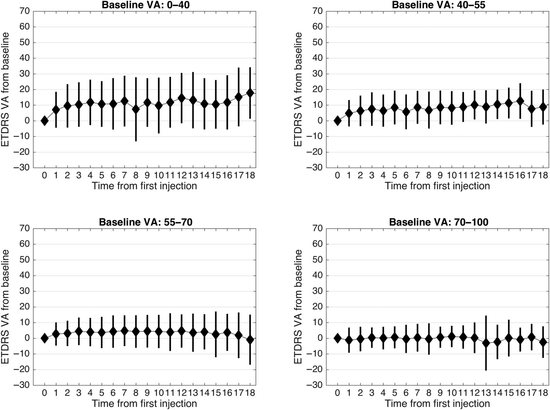
Visual acuity (VA) change in letters from baseline stratified by starting vision in ETDRS letter for treatment-naïve eyes (group 1).
Figure 2 shows the association of number of injections and VA difference between the last and first injection to the baseline VA for treatment-naïve eyes. There was no significant association between the number of injections and baseline VA, but the VA difference is negatively associated with the baseline VA.

The association of the number of injections and visual acuity difference between the last and first injection to the baseline visual acuity for the treatment-naïve eyes.
Table 1 shows the visual change and number of injections from baseline; 17.3% of eyes gained at least 15 letters. Sixty per cent of eyes were in the 0–15 letter change from baseline.
Demonstrates the visual change (letters) and number of injections (SD) from baseline for eyes receiving at least 3 injections and with a baseline visual acuity recorded within 4 weeks of the first injection during the entire follow up period
The baseline DR grade within 4 months of the first injection did not seem to influence the average VA outcomes (see online supplementary figure S2) in the first year of treatment.
Online supplementary figure S3 highlights the change in mean VA between patients with up to 1 year of follow-up (n=1136) and those with >1 year of follow-up (n=363).
Effect of baseline characteristics on VA change
The treatment effect, quantified as the VA difference between the last and first injection, is negatively associated with baseline acuity (figure 2) for treatment-naïve eyes, there was a ‘ceiling effect’ for those with better vision, who showed a reduced visual gain compared with baseline. Of note, the VA change from the baseline for eyes receiving at least three ranibizumab treatments was stratified by the baseline VA of 70–100 letters (548 eyes), 55–69 letters (813 eyes), 40–54 letters (422 eyes) and 0–39 letters (403 eyes) and is illustrated in figure 1. Eyes in the 55–70 letter group gained a mean of 5 letters at month 12 but not at month 18, and in the 40–55 letter group gained a mean of 10 letters, which was maintained at month 12 and at month 18. The poor baseline VA group (0–40 letters) gained a mean of 12 letters at month 12, but the VA results from 12 to 18 months, while showing mean gains, were highly variable. Figure 3 shows mean VA versus months since the first injection.

Upper: visual acuity (mean and SD) versus months since the first injection for all treatment groups (upper graph) and the number of eyes contributing to each data point (lower). Lower: number of eyes treated plotted against months from first injection of ranibizumab for eyes with a baseline visual acuity recorded within 1-month of the first injection.
Number of injections and visits
The frequency of ranibizumab injections at various treatment lengths is shown in online supplementary figure S4. Eyes followed-up for at least 6 months received a mean of 3.3 injections. The mean number of outpatient visits (normalised to allow comparison of one stop and two stop services as discussed in the Methods section) in the first year of follow-up was 6.9 visits.
Discussion
This study confirms that clinics using EMRs that mandate collection of nationally agreed datasets can prospectively collect large volumes of ‘real-life’ outcomes data on patient characteristics, DMO treatments and visual outcomes that can be rapidly extracted for analysis. Data were collected as part of routine clinical care and form part of the patient's contemporaneous clinical care record. One potential weakness of this study is that some centres used the EMR only for patients receiving injections and that patients apparently lost to follow-up were in fact still under review in outpatient clinics. Patients with either very good or very poor responses to therapy may have been lost to this dataset in those centres. Key missing data points that represent areas of influence in designing care pathways (eg, ethnicity data, or validated type of diabetes mellitus, VA data on non-treatment clinic visits) will be important in the future designs of ophthalmic EMRs.
In this study, there was a mean gain of five letters at 12 months after initiation of treatment when both treatment-naïve and previous treatment groups were analysed together. This is quite disappointing compared with the pivotal trials,2 ,3 ,5–7 ,9 ,13–15 which observed a mean gain of 10.6–11.1 letters at 12 months. DMO observations from smaller open-label prospective, phase IIIb studies such as PRIDE (Efficacy and Safety of Ranibizumab Alone or in Combination With Laser Photocoagulation vs. Laser Photocoagulation Alone in Proliferative Diabetic Retinopathy) (n=515), RELIGHT (Ranibizumab Treatment of Diabetic Macular Oedema With Bimonthly Monitoring After a Phase of Initial Treatment) (n=108) and RETAIN (Long-term Outcomes in Patients with Retinal Vein Occlusion Treated with Ranibizumab) (n=332) observed slightly greater VA acuity gains at >12 months ranging from four to eight letters.16 ,17 However, the gain at 12 months is similar to real-world studies16 ,18 and within the range from an analysis of several trials.19
Our findings from the UK real-world practice may reflect the more chronic nature of the disease in some patients, who had waited for access to ranibizumab or received other treatments for DMO prior to national approval; the presence of other ocular or systemic conditions that may limit the efficacy of treatment that were excluded from clinical trials or undertreatment and insufficient follow-up visits. It is also worth noting that patients were only eligible for initiating ranibizumab therapy when their macular thickness was >400 microns. This imposed threshold may have resulted in the increased chronicity of disease in our UK-based cohort. The impact of a longer duration of DMO and previous macular laser on reducing VA gains has been suggested in the outcomes from clinical trials.5 Furthermore, our findings may also reflect service delivery issues—fewer injections per eye, longer intervals between injections than clinically indicated, longer delays before initiation of treatment or recommencement of treatment after a recurrence, variability in interpretation of retreatment criteria or stopping and starting criteria.
Nonetheless, analysis of this dataset does highlight some encouraging findings. Figure 1 shows that eyes with excellent starting VA (70–100 letters) at baseline maintained good mean VA throughout the 18 months follow-up, although showed a slight negative gain (−2 letters at 18 months). This emphasises the ceiling effect of visual gain as an outcome measure and indicates that gain is not a good measure of the quality of care. In contrast, eyes with worse starting acuity achieved a greater gain in acuity. However, the final VA achieved in these patients, which is often the most important outcome for the patient, was not as good as those with a good baseline VA. This has been similarly observed in other diseases, such as age-related macular degeneration treated with ranibizumab20 and in another study of DMO outcomes.19
There were 373 eyes in the worst VA group and the reasons for the variability in vision in the second year of follow-up warrant further study. It is possible that the first-year VA results reflect resolution of macular oedema and the second-year results reflect other ocular pathologies, such diabetic macular ischaemia or poor glycaemic control. A recent study suggested that the level of diabetic control may not influence VA outcomes.9
Role for benchmarking standards
‘Translating’ the results of clinical trials into real-world clinical settings is a well-recognised problem that well-designed EMR systems can inform. In order to understand how well we are delivering care, we need to understand what can be achieved in unselected patient populations and how they differ from clinical trials.
This study represents the visual outcomes achieved in routine clinical practice in the UK prior to 2014 and provides a ‘real-world’ benchmark to compare local outcomes. It is important to note, however, that these results were obtained with fewer injections and fewer clinical visits than the pivotal studies.
An important benchmark for visual function is the proportion of patients who achieve 20/40 or better, which approximates the UK driving standard threshold. In this study, 33% of patients achieved better than or equal to 20/40 in the treated eye at 12 months. In the RISE and RIDE trials at baseline, this proportion of eyes was 19.7% and 19.2% in the control arms and 19.2% and 19.7% in the 0.5 mg ranibizumab treatment arms. At 12 months, this proportion of eyes was 34.6% and 37.8% in the control/crossover arms and 62.2% and 63.2%21 in the 0.5 mg ranibizumab arms.
Another important benchmark is the stability of vision after the maximum VA gained. Vision stability is thought to be an important outcome in other studies7 ,21 and is clearly of primary importance to patients. We suggest that VA stability is an important measure of the quality of service delivery. It is independent of baseline acuity and may therefore be used as a metric for comparisons between different population groups. In our cohort, VA gains at 6 months were stable at 18 months, except the group with the worst starting VA.
References
Footnotes
Twitter Follow David Crabb @crabblab
Collaborators UK AMD and DR EMR Users Group: Contributing Centres and Lead Clinician at each centre: Belfast Health and Social Care Trust, Professor Usha Chakravarthy; Calderdale and Huddersfield NHS Foundation Trust, Mrs Rehna Khan; Cambridge University Hospitals NHS Foundation Trust, Mr Jong Min Ong; Central Manchester University Hospitals NHS Foundation Trust, Mr Sajjad Mahmood; Frimley Park Hospital NHS Foundation Trust, Mrs Geeta Menon; Gloucestershire Hospitals NHS Foundation Trust, Mr Robert Johnston; Heart of England NHS Foundation Trust, Miss Saher Al-Husainy; Hinchingbrooke Health Care NHS Trust, Ms Toks Akerele; Hull and East Yorkshire Hospitals NHS Foundation Trust, Miss Louise Downey; Leeds Teaching Hospitals NHS Trust, Mr Martin McKibbin; Mid Yorkshire Hospitals NHS Trust, Mr Atul Varma; Moorfields Eye Hospital at Bedford Hospital, Mr Aires Lobo; Northern Devon Healthcare NHS Trust, Dr Elizabeth Wilkinson; Peterborough and Stamford Hospitals NHS Foundation Trust, Mr Alan Fitt; Sheffield Teaching Hospitals NHS Foundation Trust, Mr Christopher Brand; University Hospitals Birmingham NHS Foundation Trust, Miss Marie Tsaloumas; University Hospitals Bristol NHS Foundation Trust, Miss Clare Bailey; Warrington and Halton Hospitals NHS Foundation Trust, Miss Kaveri Mandal; Wirral University Teaching Hospital NHS Foundation Trust, Mr Vineeth Kumar; Wrightington, Wigan and Leigh NHS Foundation Trust, Mr Salim Natha.
Contributors All the authors have contributed to the planning, conduct and reporting of the work described in the article.
Funding This work was supported in part by an unrestricted research award by Novartis Pharmaceuticals. This research has received a proportion of its funding from the Department of Health's NIHR Biomedical Research Centre for Ophthalmology at Moorfields Eye Hospital and UCL Institute of Ophthalmology.
Competing interests RJ is the Medical Director of Medisoft, the Electronic Medical Record software provider from which data were extracted.
Provenance and peer review Not commissioned; externally peer reviewed.
Linked Articles
- At a glance