Article Text

Abstract
Background/aims: Glial fibrillary acidic protein (GFAP) is an established indicator of retinal stress; its expression in retinal astrocytes and Müller cells has been demonstrated to be modulated by cytokines and retinal pathology, including age related macular degeneration (AMD). This study aims to quantify the modulation of GFAP expression in retinas with drusen and atrophic AMD versus normal age matched controls.
Methods: Following a histopathological survey, 17 donor retinas were classified into four groups: drusen (n=5), geographic atrophy (GA) (n=6), aged normal (n=3), and young normal (n=3). Paramacular cryosections were immunolabelled with GFAP antibody, examined by confocal microscopy, and quantified by NIH digital image analysis. Groups were matched for potential confounding factors including age, sex, and postmortem delay.
Results: A significant increase in GFAP immunolabelling of macroglia was noted in aged normal compared with young normal retinas (p<0.04). Upregulation of GFAP immunoreactivity involving astrocytes was observed in drusen retinas compared with control retinas (p<0.03). GFAP was also upregulated in retinas with GA compared with controls (p<0.05) and in retinas with GA compared with drusen (p<0.04), both involving Müller cells. Discrete regions of GFAP upregulation in Müller cells were associated with drusen formation. In GA specimens atrophied retinal pigment epithelium (RPE) was substituted by GFAP immunoreactive Müller cell processes (gliosis).
Conclusion: This study provides a quantitative assessment of GFAP modulation in ageing and AMD affected retinas. Morphological observations were consistent with quantitative analyses indicating differential modulation of GFAP immunoreactivity in inner and outer retina. Upmodulation of GFAP in inner retina and astroglial processes was predominantly associated with drusen, while in outer retina Müller glia upmodulation of GFAP was associated with disruption of the RPE and blood-retinal barrier.
- drusen
- geographic atrophy
- macroglia
- image analysis
Statistics from Altmetric.com
The global prevalence of age related macular degeneration (AMD) continues to increase in relation to ageing of the population.1 Therapies for wet AMD are currently under development2; however, treatment is unavailable for the dry form which also represents a major cause of severe visual impairment.3
The International Age-Related Maculopathy Epidemiological Study Group defined early AMD as the presence of drusen with/without changes in retinal pigment epithelium (RPE); and late AMD as encompassing both dry AMD (geographic atrophy; GA) and wet AMD (exudative/neovascular).4 Among those with early AMD, the 5 year incidence of late AMD is estimated at 11.7%.5 The significance of small hard drusen in relation to AMD/ageing6 and the precise relation of drusen to the pathogenesis of the late stages of AMD remain to be fully defined.7
Macroglial cells in the mammalian retina (astrocytes and Müller cells) have been observed to have an active role both in normal retinal function8 and pathology.9 Glial fibrillary acidic protein (GFAP), an established marker of glia, is constitutively expressed by human retinal astrocytes.10 Upregulation of GFAP expression, an indicator of reactive gliosis, has been demonstrated in human Müller cells in response to various retinal pathologies (aberrant expression).11,12 The expression of GFAP in macroglia has also been shown to be modulated in vitro and in situ by several cytokines and growth factors.13,14
Features suggestive of early reactive gliosis have been observed associated with normal ageing of the mammalian CNS.15 Studies of the effects of ageing on human retinal macroglia are relatively few16,17; however, histological and psychophysical studies of human retinas suggest that photoreceptor abnormalities associated with ageing and AMD may share a common mechanism.18
Previous histopathological studies of AMD have focused mainly on the involvement of the outer retina including the photoreceptors, RPE, and choroid.18–20 Quantification of reactive gliosis in AMD and in normal human retinal ageing has not been extensively investigated. One study has, however, described quantitative analysis of the lateral spread of GFAP expression in Müller cells (reactive gliosis) after laser lesion in whole mounts of rabbit retinas.21 In the present study, we have quantified GFAP expression in sections of retinas with AMD using immunohistochemistry, confocal microscopy, and digital image analysis.
MATERIALS AND METHODS
Specimens
The study was performed under the tenets of the Declaration of Helsinki. Adult human eyes were obtained subsequent to informed consent through the Lions New South Wales Eye Bank. Following removal of the anterior segment and vitreous, eyecups were fixed in 2% paraformaldehyde/0.1M phosphate buffered saline (PBS; pH 7.4) and stored at 4°C.
Histopathological survey
Eyes were surveyed for pathology as follows. Macular trephines of 7 mm diameter comprising all retinal layers and choroid were taken. The neural retina was dissected free from each trephine, and the RPE-choroid portion was postfixed in 2.5% glutaraldehyde and 2% osmium tetroxide, block stained in uranyl acetate, dehydrated through graded alcohols and acetone, then embedded in Epon-Araldite resin (ProScitech, Townsville, Qld, Australia). Semithin and ultrathin sections were cut and examined by light and transmission electron microscopy for evidence of AMD. Specimens were subsequently classified into the following groups: young normal (<45 years of age), aged normal (>50 years of age), and AMD (early and late).
Cryosection preparation
Four mm trephines were taken from the superotemporal paramacular region of each eye. The trephines were rinsed several times in 0.1M PBS for 2 hours, then cryoprotected in 30% sucrose in PBS for 90 minutes. Particular care was taken to preserve the anatomical relation between the neural retina and the RPE-choroid layers. The tissues were embedded in Tissue Tek OCT Compound (Sakura Finetek, Torrance, CA, USA), snap frozen in liquid nitrogen cooled isopentane (BDH Pty Ltd, Sydney, Australia) and stored at −80°C. Cryosections of 15 μm thickness were collected on poly-l-lysine (Sigma-Aldrich Pty Ltd, Sydney, Australia), and gelatin (BDH Pty Ltd, Sydney, Australia) coated slides for immunohistochemistry.
Experimental design
We have previously demonstrated that neither postmortem delay (PMD) for up to 30 hours nor storage duration (SDu) of tissue in fixatives has significant effects on GFAP immunoreactivity in normal human adult retinas.22 Following histopathological survey, 17 donor eyes were classified into four groups and all groups were matched for PMD, SDu, and sex, and the latter three groups matched for age, as follows: young normal (n=3; mean age 27.7 (SD 5.5) years; mean PMD 17.3 (3.5) hours; mean SDu 5.7 (4.0) years), aged normal (n=3; mean age 81.7 (1.5) years; mean PMD 16.5 (4.0) hours; mean SDu 5.7 (3.8) years), early AMD (drusen; n=5; mean age 82.5 (11.2) years; mean PMD 14.5 (8.4) hours; mean SDu 5.5 (4.9) years), and late AMD (GA; n=6; mean age 79.3 (2.9) years; mean PMD 18.5 (8.2) hours; mean SDu 2.8 (1.8) years). Eyes with a known history of chronic neurological or other ocular diseases were excluded from the present study (Table 1).
Donor information
Immunohistochemistry
A standard three step indirect immunofluorescence technique was performed. Following serum blocking (10% normal donkey serum NDS/0.1M PBS) for 15 minutes, sections were incubated in polyclonal rabbit anti-cow GFAP antibody (1:5000 in PBS/2% NDS; Dako Australia, Botany, NSW, Australia) at 4°C overnight. The 1:5000 dilution was determined by titration to be the concentration of primary antibody that produced distinct GFAP labelling in astrocytes, with minimal labelling of Müller cells, in normal retinas. After rinsing with 2% fetal bovine serum (FBS)/0.1M PBS for 15 minutes, sections were incubated in biotinylated donkey anti-rabbit secondary IgG antibody (1:50 in PBS/2% NDS; Amersham Pharmacia Biotech Australia, North Ryde, Australia) for 45 minutes. After a further rinse in 0.1M PBS, sections were incubated with streptavidin conjugated Cy3 (1:100; Zymed, San Francisco, CA, USA) for 20 minutes. After rinsing in 0.1M PBS, some slides were additionally counterstained with 4 μg/ml Hoechst 33258 (Bis-benzimide; Molecular Probes Inc, Eugene, OR, USA) to show cell nuclei. The primary antibody was omitted in negative controls. After rinsing, slides were coverslipped in glycerol and examined by two independent observers using a Leitz Diaplan fluorescence microscope. Photographs were taken using the Ektachrome colour reversal film and colour slides were subsequently scanned on a Microtek ScanMaker 8700 and assembled as a composite photomicrograph using Adobe Photoshop (Version 5.0.2).
For double immunolabelling, sections were blocked in 10% normal donkey and goat serum/0.1M PBS, then incubated in polyclonal rabbit anti-cow GFAP antibody (1:5000) and mouse anti-swine vimentin (1:100) (Dako Australia, Botany, NSW, Australia) at 4°C overnight. After rinsing, specimens were incubated in biotinylated donkey anti-rabbit secondary IgG antibody (1:50; Amersham Pharmacia Biotech) for 45 minutes, rinsed in 0.1M PBS, followed by incubation in streptavidin conjugated Cy3 (1:250, Zymed) for 20 minutes. After rinsing, sections were then incubated in AlexaFluor 488 goat anti-mouse IgG conjugate (1:1000, Molecular Probes Inc). Following a further rinse, slides were coverslipped in glycerol and examined using a Leica TCS-NT scanning laser confocal microscope system.
Confocal microscopy
Following immunolabelling, cryosections were examined and images acquired using a Leica TCS-NT scanning laser confocal microscope system (Leica Microsystems, Heidelberg, Germany). Fluorochrome labelled specimens were stimulated with argon-krypton laser at 568 nm and seven optical sections of 1.5 μm thickness were collected by photomultipliers using a PLAPO 16× oil immersion objective and appropriate band pass filters. Settings including photomultiplier gain, offset, aperture, and laser power were standardised and maintained rigorously to allow comparison of GFAP labelling intensity between different specimens. Two random fields per specimen section were analysed (Fig 1A).

(A) Schematic diagrams illustrating sampling procedures for confocal microscopy and (B) “on-screen” definitions of inner and outer retinal sample zones from a merged confocal microscopy image.
Digital image analysis
GFAP immunoreactivity was quantified using digital image analysis of the confocal images. Optical sections from each field of the specimen were imported into the NIH Image program (National Institute of Health, Bethesda, MA, USA), combined, and averaged into one image. Binarisation of the images (conversion to a grey scale) was performed, allowing the computer to distinguish between areas of immunoreactivity and background.23 A standardised background elimination process was conducted (including elimination of the RPE autofluorescence), and two uniform fields in each image were subsequently defined for analysis. On the computer monitor image, a 5 mm wide band within the nerve fibre layer (NFL) and a 40 mm wide band extending from the outer limiting membrane (OLM) to the inner plexiform layer/inner nuclear layer (IPL/INL) were selected (Fig 1B); the inclusion of the defined fields in all specimens was ensured. The total field was also analysed. Segmentation of immunoreactivity was achieved using a standardised custom histogram based thresholding technique, and then subjected to particle analysis. Two parameters were recorded: (1) absolute fluorescence intensity per field, and (2) percentage area of field labelled (that is, sum of particle analyses; arbitrary units, AU). The data were then imported into Excel where the third parameter, total fluorescence intensity (absolute intensity × area; arbitrary units, AU), for each field of the specimen was calculated. The means of the latter two parameters (GFAP immunolabelling area and intensity respectively) were obtained for each group and compared.
Statistical analysis
Quantitative measurements from the four comparison groups (drusen, GA, aged normal, and young normal) were analysed by an unpaired analysis of variance (ANOVA) test, followed by the Student-Newmann-Keuls post hoc test to further evaluate the significant differences among the groups. The p values for two group comparison were obtained via the Student’s t test. All analyses were performed utilising the InStat 2.01 statistical program (GraphPad Software Systems, San Diego, CA, USA), and p<0.05 was considered statistically significant.
RESULTS
Histopathological evaluation
Histopathological survey of postmortem human eyes revealed the following features of AMD.19 RPE disturbance consisted of depigmentation, hypopigmentation or hyperpigmentation, RPE hypertrophy, hyperplasia, and atrophy. Retinas with basal laminar/linear deposit (BLD) were characterised by the presence of abnormal collagen deposition between the plasma membrane and the basal lamina of the RPE. Drusen were seen as acellular amorphous excrescences deposited between the basal lamina of the RPE and the inner collagenous layer of Bruch’s membrane. Additionally, in areas of more advanced drusen formation, thinning, depigmentation, and disruption of the overlying RPE were observed. Disciform scarring appeared as subretinal fibrovascular or fibroglial tissues, often seen replacing the RPE and associated with accumulations of leucocytes, lymphocytes, and plasma cells in the choroid (see Figs 4A–C, Penfold et al7). Geographic atrophy (GA) was characterised by gliosis associated with regions of hypopigmentation, depigmentation or apparent absence of the RPE, and/or pigment clumping; with or without photoreceptor involution and choroidal atrophy. Chronic inflammatory cells were also seen associated with atrophic lesions. Retinas classified as normal had no evidence of RPE or choroidal changes.
Retinas included in the current study were noted to have the following features: moderate RPE changes were observed in 2/5 drusen retinas (Figs 3A and B). In retinas classified as GA, pigment clumping was observed in 5/6 specimens (Figs 3D, 4A–C, and E), drusen were present in 2/6 retinas (Figs 3D and 4B), and gliotic lesions were evident in 2/6 specimens (Figs 4C and E).
GFAP immunoreactivity in normal retinas
GFAP immunolabelling in all young and aged normal specimens (6/6) was found predominantly in astrocytes in the NFL ganglion cell layer (GCL) (inner retina) (Figs 3E and 4F). Occasionally, Müller cell processes extending between the ILM and OLM were noted to display low level GFAP immunolabelling in one young normal and two aged normal retinas (Fig 3E).
GFAP immunoreactivity in AMD
GFAP immunolabelling in astrocytes appeared more intense in all AMD specimens compared with normal controls. Increased GFAP labelling in Müller cells (outer retina) was observed in 2/5 drusen (Figs 2B and Cv 2E) and 6/6 GA (Figs 3D and 4A–E v 4F) specimens compared with controls. Note that outer retinal GFAP expression was seen predominantly in the outer Müller cell processes (OLM-IPL/INL) and only occasionally in the entire Müller cell cytoplasm (OLM-ILM) as in Figure 4D. Interestingly, focal areas of intense upregulation of outer retinal GFAP expression were observed associated with regions of drusen formation, in three specimens—one drusen associated retina (Fig 3C) and both GA retinas with drusen (Figs 3D and 4B). Widespread outer retinal GFAP expression was noted in all other GA specimens (Fig 4A, C, and E). In addition, regions of intensely GFAP immunoreactive gliosis were observed in two GA specimens where atrophied RPE was substituted by GFAP immunoreactive Müller cell processes overlying Bruch’s membrane (Figs 4C and E). Immunolabelling was absent in the negative control (Fig 3F). Selected sections were counterstained with Hoechst (bis-benzimide) nuclear stain in order to show the retinal layers and GFAP immunolabelled glia (Figs 2A–C).

Hoechst counterstained GFAP immunolabelled normal (A), drusen (B), and GA (C) specimens showing retinal layers. In (D–F), an example of a GA specimen double immunolabelled using GFAP (red) and vimentin (green) is seen. The predominant expression of GFAP in astrocytes and relative paucity of expression in Müller cell end feet is illustrated in (D). The predominant expression of vimentin in Müller cell end feet and relative paucity of expression in astrocytes is seen in (E). By overlaying (D) and (E), a small degree of co-expression of GFAP and vimentin in Muller cell end feet is seen in (F). However, a distinct pattern of GFAP expression is seen predominantly in astrocytes versus vimentin, predominantly observed in Muller cell end feet (F). NFL = nerve fibre layer; INL = inner nuclear layer; ONL = outer nuclear layer; PRL = photoreceptor layer; RPE = retinal pigment epithelium; McE = Müller cell end feet; E = epithelium; Bv = blood vessel; G = gliotic lesion.

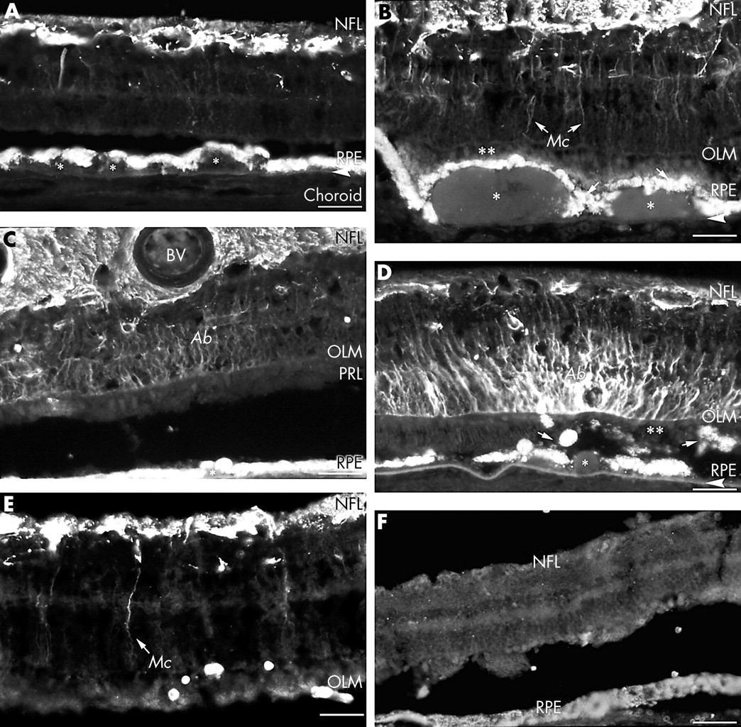
GFAP immunofluorescence labelling of cryosections of retinas classified as drusen (A–C), GA (D), or control (E and F). Upregulation of GFAP immunoreactivity is seen in astrocytes (in the nerve fibre layer, NFL) in all AMD retinas compared with the normal control. Note that the retinal pigment epithelium (RPE) displays autofluorescence in all micrographs. (A) Illustrates multiple drusen (*) above Bruch’s membrane (arrowhead), RPE disturbance, and intense GFAP immunoreactivity in astrocytes; although minimal GFAP immunoreactivity is seen in Müller cells (donor age 88 years). (B) Demonstrates large drusen (*) above Bruch’s membrane (arrowhead). Note that overlying RPE disturbance (arrows) and photoreceptor involution (**) are associated with enhanced immunoreactivity in Müller cells (Mc) (donor age 66 years). OLM = outer limiting membrane. (C) Illustrates focal enhanced immunoreactivity in Müller cells (Ab) in a drusen specimen. Note the large blood vessel (BV) in the NFL (donor age 93 years). PRL = photoreceptor layer. (D) A specimen with GA demonstrates focal enhanced immunoreactivity in Müller cells (Ab) in regional association with drusen formation (*), pigment clumping (arrows), and photoreceptor involution (**) (donor age 78 years). (E) Young normal retina showing much reduced immunoreactivity in the NFL. Note normal RPE and absence of Müller cell labelling (donor age 22 years). (F) Negative control showing RPE autofluorescence and an absence of immunolabelling in the neural retina. Scale bars = 125 μm.
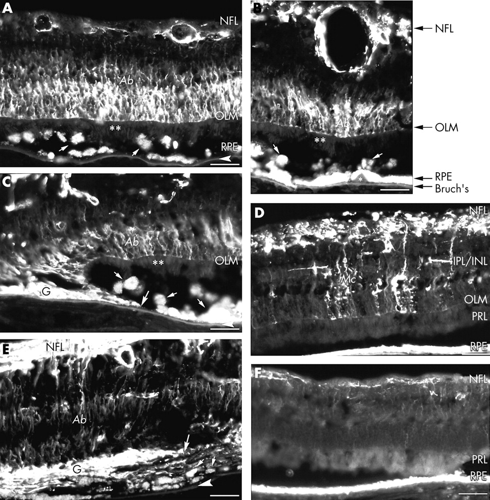
GFAP immunofluorescence labelling of cryosections of retinas classified as GA (A–E) or normal (F). Significantly increased GFAP immunoreactivity was noted in both astrocytes (that is, inner retinal expression; in NFL) and Müller cells (that is, outer retinal expression, aberrant, Ab) in all GA specimens, compared with the normal control. The negative control is shown in (F). Note that GFAP expression in Müller cells mostly extends from the OLM to the inner plexiform layer/inner nuclear layer (IPL/INL), and only occasionally extends between the OLM and the ILM. Pigment clumping/RPE disturbance (small arrows) and photoreceptor involution (**) are also visible in (A–C, and E). RPE displays autofluorescence in all micrographs. (A) Shows extensive outer retinal GFAP immunoreactivity (Ab) in Müller cells associated with pigment clumping (small arrows) and photoreceptor involution (**). Bruch’s membrane = arrowhead (donor age 78 years). (B) illustrates focal enhanced outer retinal GFAP expression (Ab) in direct association with drusen formation (*) above Bruch’s membrane (arrowhead), pigment clumping (small arrows), and photoreceptor involution (**) (donor age 78 years). (C) Further illustrates the same features as in (B) (Ab, arrows, and **). In addition, a gliotic lesion (G; edge denoted by long arrow) with enhanced immunoreactivity within Müller cells is seen overlying Bruch’s membrane (arrowhead); RPE cells are absent (donor age 78 years). (D) Shows enhanced GFAP immunoreactivity in astrocytes and Müller cells (Mc). Occasional horizontal Müller cell processes are seen in the inner plexiform layer/inner nuclear layer (IPL/INL) (donor age 83 years). PRL = photoreceptor layer. (E) Demonstrates increased inner retinal and outer retinal (Ab) immunoreactivity. An immunoreactive gliotic lesion (G; edge denoted by long arrow) is seen associated with RPE disturbance (small arrows) and complete absence of photoreceptors (donor age 75 years). (F) Aged normal retina showing reduced GFAP immunoreactivity in the NFL; normal RPE and an absence of Müller cell labelling (donor age 80 years). Scale bars = 125 μm.
The predominant expression of GFAP in astrocytes and relative paucity of expression in Müller cell end feet is illustrated in Figures 2D–F. By overlaying Figures 2D and E, a small degree of GFAP and vimentin co-expression is visible in Muller cell end feet (Fig 2F). However, the distinct pattern of GFAP expression seen predominantly in astrocytes v vimentin, predominantly visible in Muller cell end feet, is apparent.
Quantitative assessment
Following quantitative analyses, GFAP immunolabelling intensity (Fig 5A–C) and area (Fig 5D–F) for the three regions of expression (inner retinal, outer retinal, and total) in each histopathological group (drusen, GA, young normal, and aged normal) were plotted. ANOVA statistical analysis revealed significant increases in GFAP immunoreactivity (both labelling intensity and area) in AMD affected retinas compared with control specimens for the three regions of expression (inner retinal, outer retinal, and total) (Figs 5B, C and D–F; p<0.05), except for inner retinal GFAP labelling intensity (Fig 5A; p=0.0688).

Graphs showing GFAP immunoreactivity in four histopathological groups—drusen (DRU), geographic atrophy (GA), aged normal (AN), and young normal (YN) controls. GFAP immunoreactivity was assessed for two factors: fluorescence immunolabelling intensity (A–C) and unit area labelled (D–F). Three regions of GFAP expression were analysed: (A and D) Inner retina astrocytes in the NFL/GCL. (B and E) Outer retina Müller cells between OLM and IPL. (C and F) Total— including inner retina and outer retina. p Values shown in each graph are for comparison of the four histopathological groups via an unpaired ANOVA test.
The Student-Newmann-Keuls post hoc test was performed to further evaluate significant differences among the groups and p values obtained using the Student’s t test were tabulated for each comparison group (Table 2). Note that significant increases in inner retinal and total GFAP immunoreactivity (both labelling intensity and area) were found for drusen v controls (Table 2; Figs 5A, C, D, and F); in outer retinal and total immunoreactivity for GA v controls (Table 2; Figs 5B, C, E, and F); and in outer retinal immunoreactivity for GA v drusen (Table 2; Figs 5B and E). Increased total GFAP labelling area, but not intensity, was observed in aged normal compared with young normal retinas (p=0.0383) (Table 2; Fig 5F).
p Values* for comparison of GFAP immunoreactivity† between different histopathological groups for the three regions analysed
When inner retinal and outer retinal GFAP immunoreactivities were compared, similar levels were found in retinas with GA (labelling intensity p=0.9539; labelling area p=0.5931). However, outer retinal expression was significantly lower than inner retinal expression in the drusen group (intensity p=0.0009; area p=0.0006) (Figs 5Av B and 5D v E).
DISCUSSION
In the normal mammalian retina GFAP is recognised as a primary marker of astrocytes within the NFL and GCL,10 while it is marginally detectable in Müller cells24 which extend radially from the OLM to the ILM.25 Consistent with these observations, GFAP expression in astrocytes is referred to as inner retinal and expression in Müller cells as outer retinal, as defined above.
Numerous studies have reported reactive gliosis involving Müller cells in vertebrate retinas in response to various pathologies including mechanical injury,26 retinal detachment,27 diabetic retinopathy,12 glaucoma,28 retinal ischaemia,29 and retinal degeneration.30 Increased GFAP expression in macroglia has also been described in retinas with AMD.16,17,20 The current study further provides a quantitative comparison of GFAP immunoreactivity, demonstrating differential modulation of GFAP expression in retinal macroglia in aged v young normal retinas, and in early v dry AMD. The significance and possible implications of these observations are discussed as follows.
GFAP immunoreactivity in aged human retina
Aged human CNS including the retina has been suggested to display early features of reactive gliosis (reviewed Cotrina and Nedergaard15). In particular, astrocytes in the aged human retina have been described to undergo morphological changes including hypertrophy and increased density of intermediate filaments, displaying increased GFAP immunoreactivity.16,17 Increased GFAP expression has also been demonstrated in Müller cells in the aged human retina.16 In the present study we observed significant increases in GFAP immunolabelling in aged normal compared with young normal retinas in both astrocytes and Müller cells, consistent with the above observations. Such an age associated response of astrocytes (and perhaps Müller cells) has been proposed to exert a protective action against oxidative stress,31,32 and to parallel human neurodegenerative disorders,15 although the exact mechanisms remain unknown. Further, some studies have suggested that retinal ageing and AMD may be part of a continuous process of deterioration.33,34
GFAP immunoreactivity in early v dry AMD
In the present study, significant increases in GFAP expression were observed in astrocytes in drusen retinas, and in Müller cells in GA. This differential modulation implies that astrocytes and Müller cells may have specific roles in the pathogenesis of AMD, rather than displaying a generalised response to retinal stress. Furthermore, consistent with our previous observations16 and a recent study of late AMD (dry and wet),20 we have observed focal upregulation of GFAP expression in Müller cells, directly associated with areas of drusen formation (Fig 2D). Widespread upregulation of Müller cell GFAP expression was observed in the GA group in the present study. Overall, GFAP modulation was related to drusen and geographic atrophy, such that GA was associated with outer retinal and drusen with inner retinal modulation of GFAP.
Histopathological studies have reported a close association between drusen and increased risk of dry AMD6,35 (reviewed by Penfold et al7). Taken together, these observations indicate that reactive gliosis involving astrocytes and Müller cells represents an early response in dry AMD.
GFAP immunoreactivity in retinas with drusen
The significance of GFAP upregulation in retinal pathology, including AMD, remains unclear.12 Müller cells are closely coupled structurally and metabolically to photoreceptors,36 and changes in Müller cells have been shown to precede photoreceptor degeneration.37 In addition, Müller cells express vasomodulatory substances including nitric oxide synthase8 and endothelin,38 and produce molecules that confer barrier properties to vascular endothelium,39 thus playing a part akin to astrocytes in regulation of the blood-retinal barrier.40,41 Ramírez10 refers to the generally similar characteristics of astrocytes in AMD and normal old individuals while referring to extremely high GFAP immunoreactivity in individual cells, without distinguishing between astrocytic and Müller glia. The distinct features of GFAP immunoreactivity observed in the drusen retinas in the present study (that is, increased GFAP immunoreactivity in astrocytes and focal GFAP immunoreactivity in Müller cells associated with drusen formation) may implicate macroglial changes and blood-retinal barrier breakdown in early AMD. However, whether these changes are causal or consequential in the pathogenesis of AMD could not be ascertained by the current study.
The role of cytokine modulation
GFAP expression in macroglia has been shown to be modulated, in vitro and in situ, by several cytokines and growth factors including basic fibroblast growth factor (bFGF),13,14 ciliary neurotrophic factor (CNTF),42 transforming growth factor β,14,43 interleukin 1, interleukin 6, and interferon γ.44,45 In situ GFAP upregulation in mammalian Müller cells in response to retinal pathology has been suggested to be mediated by growth factors.13,46 Additionally, both bFGF and CNTF, endogenously expressed by the retina, have been shown to have potent protective/rescue effects on photoreceptors against a variety of retinal insults.47 Pigment epithelium derived factor, a glycoprotein normally secreted by RPE cells,48 has also been demonstrated to have neuroprotective49 and Müller cell gliosupportive50 actions.
GFAP immunoreactivity in retinas with GA
In GA specimens in the current study, atrophied RPE was substituted for by GFAP immunoreactive Müller cell processes (gliosis), indicating that breakdown of the outer blood-retinal barrier (the RPE) may be involved in the aetiology of AMD (Figs 3C and E). Gliosis has been suggested to be important for the protection and repair of neurons in CNS pathology.51 Similarly, reactive macroglia in AMD as observed in the present study, may also represent a protective response, perhaps by secreting/inducing barrier active factors such as bFGF,52 enhancing Müller cell gliotic response, and “sealing” the outer blood-retinal barrier.
The present study establishes a method for quantification and analysis of GFAP immunoreactivity in AMD and aged human retina. It adds to the histopathological characterisation of the pathogenesis of AMD, indicating that blood-retinal barrier breakdown may be involved in the aetiology of AMD. The differential modulation of GFAP immunoreactivity in astrocytes and Müller cells suggests that macroglia influence the pathogenesis of both early and late AMD.
Acknowledgments
The authors acknowledge the support provided by the staff of the Lions NSW Eye Bank and Dr Li Wen; and Ms Diana van Driel, for expert assistance with histology and electron microscopy. Dr Michele Madigan was supported by Sydney Foundation for Medical Research and Dr Philip Penfold was supported by Retina Australia.