Article Text

Abstract
Purpose To quantitate lens nuclear opacity using long-range swept-source optical coherence tomography (SS-OCT) images and to evaluate the correlation of this method to Lens Opacities Classification System III (LOCS III) and a Scheimpflug imaging-based grading system (Pentacam Nuclear Stage function; PNS).
Methods This study enrolled 120 participants (120 eyes) with age-related nuclear cataracts. The best-corrected visual acuity (BCVA), LOCS III nuclear opalescence (NO) and nuclear colour (NC) were obtained. The nuclear density measured using PNS function (NDPNS) was recorded. Three successive series of long-range SS-OCT images were captured, and the nuclear region was analysed using ImageJ (NIH, Bethesda, Maryland, USA) to generate SS-OCT image-based nuclear density (NDSS-OCT). The repeatability of NDSS-OCT measurement was evaluated using within-subject coefficient of variation (CVw) and intraclass correlation coefficient (ICC). Correlations of NDSS-OCT with NO and NC, BCVA and NDPNS were analysed. According to the integrity of nucleus imaged by Pentacam, patients were divided into two groups, and the parameters were compared between groups.
Results The CVw and ICC for NDSS-OCT measurement were 1.5 % and 0.994, respectively. The NDSS-OCT significantly correlated with NO (r=0.831), NC (r=0.873), BCVA (r=0.655) and NDPNS (r=0.891). The NDSS-OCT, NO and NC, and BCVA were significantly different between the two groups.
Conclusion Lens nuclear opacity quantitation using long-range SS-OCT images was repeatable and correlated well with LOCS III and PNS function. The Scheimpflug principle had a limitation in imaging dense nucleus. Long-range SS-OCT seems more promising for objectively and quantitatively assessing lens nuclear opacity.
- lens and zonules
- imaging
Statistics from Altmetric.com
Introduction
Appropriately evaluating lens nuclear opacity is vital for the documentation of the progress of nuclear cataract,1 as well as the prediction of ultrasound (US) energy expense during phacoemulsification, since the nucleus is the main part of lens that requires US energy for extraction.2
Clinically, light scattering caused by the lens can be used to assess the extent of its opacities. According to the direction of scattering, the methodology is generally divided into two categories: the approaches for forward scattering analysis and for backward scattering analysis.3 The former category include the compensation-comparison psychophysical method that requires the cooperation of participants in a subjective manner4 and the double-pass system that provides the objective measurement.5 6 They detect the intensity of straylight by analysing the retinal image quality. Previous studies have investigated their efficacy in evaluating lens opacifications5–8 and in acting as the decision-making tool for surgery.9 10
However, the backscattering was typically analysed using anterior segment imaging techniques. A commonly used subjective method is Lens Opacities Classification System III (LOCS III),11 which is currently thought of as the golden standard. In this process, the features of lens opacities should be recorded by an observer with slit-lamp photography first and compared with those described on the standard reference pictures later to estimate the type and severity of cataract. However, the examiner’s proficiency in performing this grading system, in addition to the slit-lamp settings, may have an influence on its reproducibility.12 13
Alternatively, the anterior segment image analysis based on tomography systems, such as Scheimpflug principle and optical coherence tomography (OCT), was able to quantitate the magnitude of backscatter to objectively assess lens opacifications, particularly the nuclear opacity.1 3 14–21 Pentacam (Oculus, Wetzlar, Germany) is a three-dimensional anterior segment tomographer using Scheimpflug principle. Its built-in function, Pentacam Nuclear Staging (PNS), is a densitometry for quantitating nuclear opacity using Scheimpflug images captured by the device.17
A few studies reported the nuclear opacity measurements using OCT images.18–21 A novel long-range swept-source OCT (SS-OCT) biometer, IOLMaster 700 (Carl Zeiss Meditec AG, Jena, Germany), was recently launched. It is able to obtain the B-scan image of full eye along the visual axis, which was used to quantitate the whole lens opacity by Panthier et al.22 More recently, de Castro et al 21 depicted the properties of lens opacifications using a long-range SS-OCT laboratory prototype and found a correlation between the nuclear opacity quantitated using its image and LOCS III nuclear opalescence (NO) score in 16 cataractous patients. Overall, the investigation on lens nuclear opacity measurement with long-range SS-OCT still remains rare. A larger sample size, as well as the assessment of measurement reliability, is needed to evaluate the feasibility of this method in clinic. Moreover, all previous studies using OCT devices employed one single B-scan image,18–21 which meant that only the nuclear morphology on a certain meridian was analysed, and could led to an absence of a global feature of opacity.
In this work, we focused mainly on the anterior segment imaging techniques for backscattering analyses and aimed to appraise the validity of nuclear opacity quantitation using multiple long-range SS-OCT images by evaluating its reliability and by assessing the correlation of this method to LOCS III nuclear grading system and PNS function in nuclear cataract.
Methods
Subjects
One hundred and twenty eyes from 120 patients with age-related nuclear cataracts were enrolled in this prospective study. The research protocol adhered to the tenets of the Declaration of Helsinki. All participants had submitted written informed consent.
All subjects underwent complete ophthalmic examinations, including uncorrected distance visual acuity and best-corrected visual acuity (BCVA), non-contact tonometry (TX-F; Cannon, Tokyo, Japan), slit-lamp microscopy and dilated fundus examination.
Patients with any history of ocular trauma, previous ocular surgery and laser treatment, and ocular diseases other than age-related nuclear cataract were excluded.
LOCS III nuclear scoring
Affected eyes were maximally dilated with eye-drops at a concentration of tropicamide 0.5% and phenylephrine hydrochloride 0.5%. The same experienced ophthalmologist (ZLL) examined all the eyes and scored the lens NO and nuclear colour (NC), respectively, on a scale of 0.1–6.9 according to LOCS III protocol.
Lens nuclear opacity quantitation using long-range SS-OCT
IOLMaster 700, a long-range SS-OCT biometer, is capable of generating six B-scan images of full eye, whose meridians were, respectively, at 0°, 30°, 60°, 90°, 120° and 150°. It uses a tunable laser source operating a light beam at a centre wavelength of 1055 nm and achieves a maximal scan depth of 44 mm and an axial resolution of 22 µm in tissue at a speed of 2000 A-scans per second. The software at version 1.5 installed on the system was applied in current study.
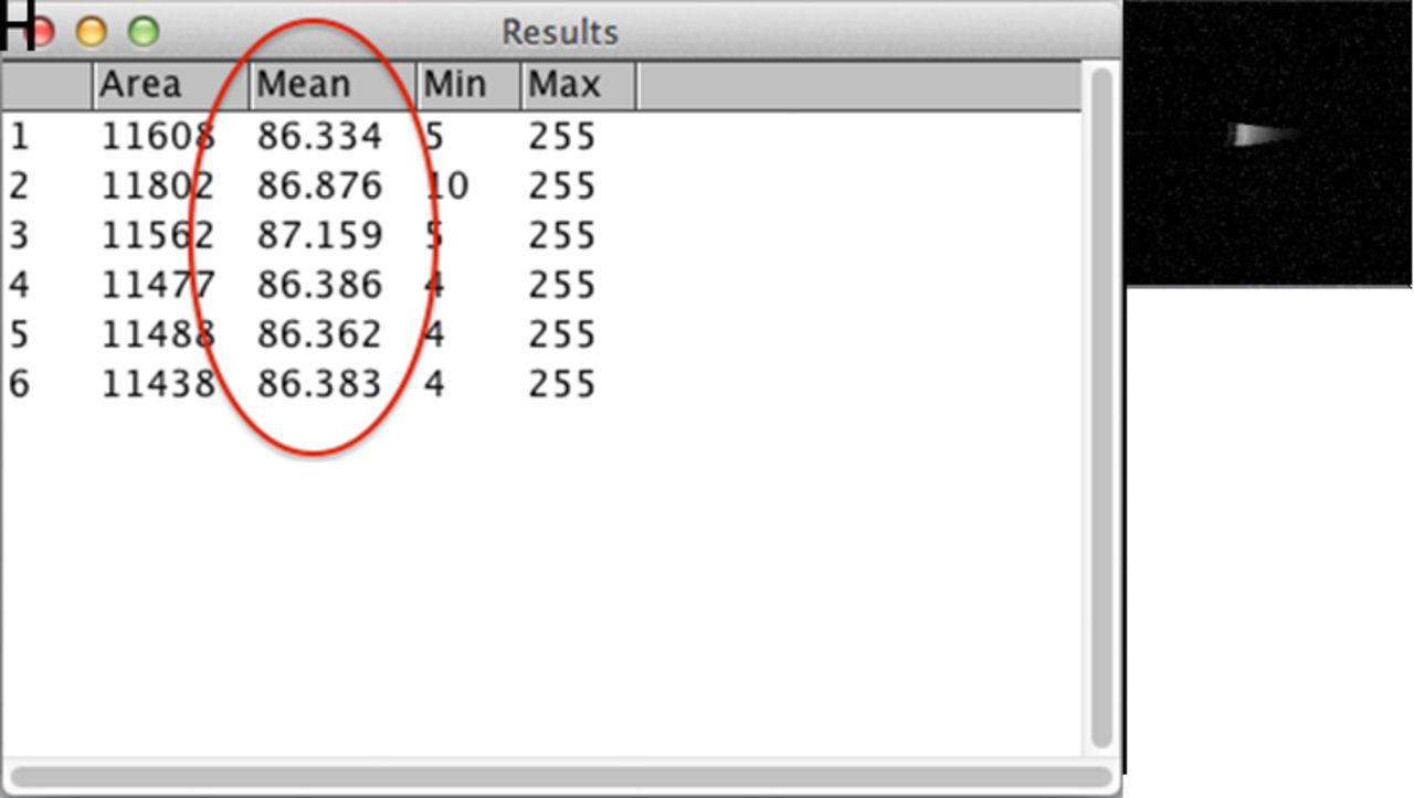
All long-range SS-OCT B-scan images were processed and analysed using ImageJ software (available at http://rsb.info.nih.gov/ij; National Institutes of Health, Bethesda, Maryland, USA).23 The entire region of lens nucleus was encompassed, and the density was measured in pixel intensity units on a scale of 0 (pure black) to 255 (pure white). For each eye, the SS-OCT nuclear density was defined as the average of the six readings derived from the corresponding images, as shown in figure 1.

The long-range SS-OCT images whose meridians were located at 0 ̊, 30 ̊, 60 ̊, 90 ̊, 120 ̊, 150 ̊, respectively (A–F), with the entire region of nucleus delineated using ImageJ software (NationalInstitutes of Health, Bethesda, Maryland, USA), the toolbar of ImageJ software (G), the outcomes of ImageJsoftware for nuclear density measurement using long-range SS-OCT images (H) and a digital slit-lampphotograph for the same lens (I). SS-OCT, swept-source optical coherence tomography.
SS-OCT nuclear density measurement procedure
After the pupil dilated, patients were seated in a dim room with chin on the chinrest, forehead against the forehead bar and instructed to fixate the fixation point. For each eye, three consecutive scans with IOLMaster 700 were performed by the same operator (DC). To assure the independence of successive measurements, patients were asked to move away their head from the chinrest, and the scan unit was thoroughly retracted after each scan.
The SS-OCT nuclear density quantitation was conducted by a masked observer (SJL) using ImageJ software. The three SS-OCT nuclear density readings for each eye were used to assess the repeatability. The average of these three outcomes was calculated for correlation analysis.
Lens nuclear opacity quantitation using PNS function
Afterwards, Pentacam HR was used to acquire the Scheimpflug images of the lens, and the average density reading for the entire nucleus provided by PNS function was defined as Pentacam nuclear density, ranging from 0% to 100% (figure 2A). The scan was thought acceptable if the ‘QS’ index showed ‘OK’.

The Pentacam Nuclear Stage user interface for density analysis (A), and the Scheimpflug image where the lens nucleus failed to be entirely captured by Pentacam HR (B).
Comparison of parameters between Pentacam group and Pentacam N/A group
In the course of Pentacam nuclear density measurement, we noticed that some of lens nuclei failed to be completely acquired with Pentacam HR (figure 2B). The participants whose nuclei were entirely obtained using Pentacam HR were assigned to Pentacam group (group 1), whereas subjects whose nuclei were not completely captured by Pentacam HR were allocated to Pentacam N/A group (group 2), where we stipulated that Pentacam nuclear density was not available. The differences in parameters, including BCVA, LOCS III NO and NC scores and SS-OCT nuclear density, between the two groups were investigated.
Statistical analysis
All statistical analyses were performed using SPSS software for Mac V.22 and Microsoft Office Excel for Mac (Microsoft Corp, Redmond, Washington, USA). A p value less than or equal to 0.05 was considered statistically significant. Data were expressed as the mean±SD or median (quartile range) depending on the normality of distribution. The data, apart from the SS-OCT nuclear density, were non-normally distributed, as checked with Kolmogorov-Smirnov test.
To evaluate the repeatability for SS-OCT Nuclear Density measurement, within-subject SD (Sw), coefficient of repeatability (CR), within-subject coefficient of variation (CVw) and intraclass correlation coefficient (ICC) were computed for the three successive measurements. The Sw was defined as the square root of within-subject mean square of error produced by one-way analysis of variance. The CR was equal to Sw times 2.77, which provided an interval within which 95% of the differences between measurements were expected to lie.24 The CVw was calculated as the Sw divided by overall mean. The lower the CVw value, the superior the repeatability was. The closer the ICC is to 1, the better the consistency for the consecutive measurements.
The correlations between SS-OCT nuclear density, BCVA (logarithmic minimal angle resolution (logMAR)), LOCS III NO and NC, and Pentacam nuclear density were analysed using Spearman correlation analysis. In addition, Mann-Whitney U test was used to compare parameters between group 1 and group 2.
Results
The study recruited 120 participants (70 men and 50 women). The mean age was 73.5±9.8 years. Only one eye of each participant was selected randomly, resulting in a data set of 62 right eyes and 58 left eyes for the analysis. The distribution of the LOCS III nuclear grading (NO and NC) scores is described in table 1.
Distribution of Lens Opacities Classification III nuclear opalescence (NO) score and nuclear colour (NC) score (n=120)
The SS-OCT nuclear density ranged from 47.3 to 121.1 with a mean of 84.0±16.6. The Sw, CR, CVw and ICC were, respectively, 1.29, 3.57, 1.5% and 0.994 (95% CI 0.992 to 0.996) for repeatability of SS-OCT nuclear density measurement.
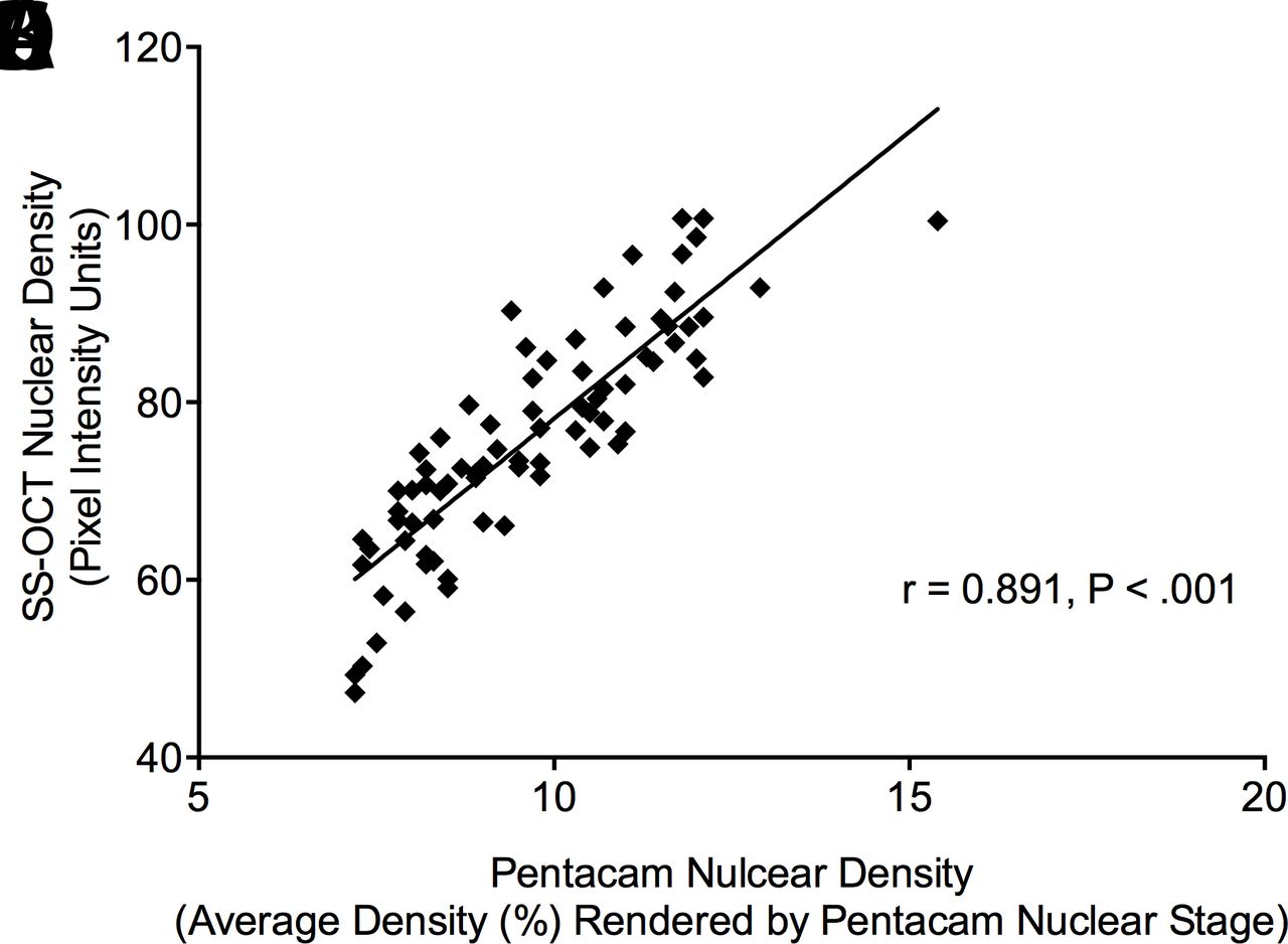
Table 2 shows the correlation coefficients between SS-OCT nuclear density and NO and NC, and BCVA. Figure 3A–C show the correlations of SS-OCT nuclear density with NO (r=0.831, p<0.001), NC (r=0.873, p<0.001) and BCVA (r=0.655, p<0.001).

The SS-OCT nuclear density, quantitated for the entire region of nucleus using long-range SS-OCTimages and ImageJ software measured in pixel intensity units, correlated with Lens Opacities ClassificationSystem (LOCS) III nuclear opalescence (NO; r=0.831, p<0.001; A), nuclear colour (NC; r=0.873, p<0.001; B), logarithmic minimal angle resolution (logMAR) best-corrected visual acuity (BCVA; r=0.655, p<0.001; C) and Pentacam nuclear density defined as average density (%) for the entire nuclear region rendered by Pentacam Nuclear Stage function (r=0.891, p<0.001; D). SS-OCT, swept-source optical coherence tomography.
Correlations of nuclear density measured using long-range swept-source optical coherence tomography image combined with ImageJ software (NDSS-OCT), nuclear density measured using Pentacam Nuclear Stage function (NDPNS), respectively, with Lens Opacities Classification System III (LOCS III) nuclear grading scores, including nuclear opalescence (NO) and nuclear colour (NC) and logarithmic minimal angle resolution (logMAR) best-corrected visual acuity (BCVA)
Of 120 eyes, 77 (64.2%) eyes were allocated to group 1 where Pentacam nuclear density was acquirable and ranged from 7.2% to 15.4% with a median of 9.7% (8.2%, 11.1%). Table 2 also shows the correlations of Pentacam nuclear density with NO, NC, BCVA and SS-OCT nuclear density. The SS-OCT nuclear density correlated strongly with Pentacam nulcear density (r=0.891, p<0.001; figure 3D).
The differences on parameters between group 1 and group 2, including BCVA (logMAR; 0.4 (0.2, 0.7) vs 0.8 (0.5, 1.3)), NO (3.0 (2.5, 3.6) vs 4.0 (3.5, 5.0)), NC (3.2 (2.8, 3.9) vs 4.7 (4.1, 5.1)) and SS-OCT nuclear density (75.3 (66.8, 85.0) vs 99.9 (89.5, 107.1)), were statistically significant (p<0.001).
Discussion
A reliable quantitative analysis system for nuclear opacity can be useful in multicentre clinical research. In addition, it could potentially help the surgeon configure the phacoemulsification instrument with reasonable settings according to the severity of nuclear opacity to optimise the efficiency and improve the safety of surgery. In this study, we concentrated on the cataractous nucleus-induced backscatter intensity rendered by the long-range SS-OCT images and used ImageJ software to determine its feasibility and advantage in characterising the degree of nuclear opacity. The low CVw (1.5%) and high ICC (0.994) indicated a high repeatability for nuclear opacity quantitation using long-range SS-OCT images.
The region of entire lens nucleus was selected for the density analysis in our study. This zone of nucleus was also used in LOCS III grading system,11 as well as nuclear density measurements using time-domain OCT (TD-OCT) images and spectral-domain OCT (SD-OCT) images.18 19 Differently, Makhotkina et al 20 defined a rectangular region of interest (ROI) on the anterior segment SS-OCT (AS-SS-OCT) image. The ROI selection method allowed the observer to choose an arbitrary area and was also used in several studies that employed Pentacam Scheimpflug images to evaluate the nuclear density.25 26 By contrast with the ROI, the entire region selection method has an advantage in portraying the morphological characteristics of the whole nucleus. Moreover, it theoretically diminishes the subjectivity of observer-defined action owing to the identifiable contour of lens nucleus.
There were strong correlations of SS-OCT nuclear density with LOCS III NO (r=0.831, p<0.001) and NC (r=0.873, p<0.001). Lower correlations of nuclear density indices based on other OCT images with LOCS III nuclear grading scores were reported.18 19 Makhotkina et al 20 quantitated the nuclear density using AS-SS-OCT image, which showed an area of highest sensitivity located in the posterior half of nucleus, where they decided to define a rectangular ROI, and the AS-SS-OCT image-based nuclear density showed lower correlations with NO (r=0.55, p<0.001) and NC (r=0.57, p<0.001), as compared with the SS-OCT nuclear density.20 The reason for this discrepancy may be the ROI they selected. The small size of ROI made AS-SS-OCT image-based nuclear density less representative for the entire nuclear opacity, which was actually limited by the scan mode of AS-SS-OCT. With regard to the long-range SS-OCT, the entire nucleus can be sharply focused, and it was fundamental to the selection of entire nuclear region for a comprehensive nuclear density assessment.
A custom-built long-range SS-OCT prototype was recently demonstrated by de Castro et al.21 The nuclear density evaluated using the image of this device showed a lower correlation with LOCS III NO (r2=0.39, p<0.005), as compared with our result (r=0.831, p<0.001). The recruitment criterion may probably be the primary contributing factor to this difference. The eyes with pure nuclear cataracts were included in present research, while mixed types of cataracts were recruited by de Castro et al.21 As was shown in their paper, both cortical and posterior subcapsular opacities could be clearly visible on the long-range SS-OCT images, which indicated that the opacifications enhanced the light reflection where they existed. It may have an impact on the intensity of light passing through the lens nucleus, and as a result, the OCT signal would alter in the nuclear region, yielding a measured value of OCT image-based nuclear density deviating from its actual outcome.
The SS-OCT nuclear density showed a significant correlation with BCVA (r=0.655, p<0.001). In contrast, Wong et al 18 found the correlation between TD-OCT image-based nuclear density and BCVA was not statistically significant. Also, Kim et al 19 revealed there was no significant correlation of SD-OCT image-based nuclear density with BCVA. However, only 55 and 47 eyes, respectively, were enrolled by Wong et al 18 and Kim et al.19 The relatively small sample size might be responsible for the insignificant correlations in their studies.
In group 1 where Pentacam achieved the imaging to the entire nucleus, Pentacam nuclear density was obtainable and showed good correlations with LOCS III NO score and NC score and BCVA. This was consistent with the previous studies.10 25 Furthermore, there was strong correlation between Pentacam nuclear density and SS-OCT nuclear density (r=0.891, p<0.001), which suggested an excellent consistency between these two approaches. Similarly, Panthier et al 22 generated a high correlation coefficient between the whole lens density quantitated using long-range SS-OCT and PNS function (r2=0.75, p<0.01). However, the correlation was weak between the whole lens density and LOCS III NO score (r=0.20, p=0.04), indicating that the whole lens density was not an ideal surrogate for the nuclear opacity.22 Makhotkina et al 20 reported a lower correlation between AS-SS-OCT image-based nuclear density and PNS-based nuclear density (r=0.54, p<0.01). This may be because the size and position of ROIs were different for Pentacam Scheimpflug image and AS-SS-OCT image, whereas in our study, the entire region of nucleus was delineated for both SS-OCT nuclear density and Pentacam nuclear density measurements.
As expected, we observed the significantly higher SS-OCT nuclear density, LOCS III NO and NC score and BCVA (logMAR) in group 2 than in group 1, suggesting that Pentacam failed to provide the Scheimpflug image of entirety of nucleus among patients who suffered severer nuclear cataract. Magalhães et al 27 revealed that, when LOCS III NO score was higher than 6.0, the correlation was statistically insignificant between NO and the nuclear density measured using PNS funcion and concluded that PNS function lost efficacy because the blue light Pentacam used was obstructed by the dense nucleus. In comparison with Pentacam, the long-range SS-OCT applies the near-infrared light with 1055 nm centre wavelength and reduces the attenuation caused by cataractous nucleus and therefore yields a deeper tissue penetration,28 competent in providing a B-scan image of nucleus even in the advanced nuclear cataract.
The long-range SS-OCT is a high-speed imaging system, capable of providing a sharply focused cross-sectional image for entire lens nucleus. However, there were two image artefacts that might influence the OCT image-based nuclear density measurement. One was the specular reflection that caused the localised signal enhancement within the nuclear region. Although its impact had been minimised by the entire nuclear region selection method we applied, it would still more or less increase the measured value of nuclear density. The other was the loss of OCT signal strength artefact induced by the aberration. The backscattered OCT beam would not be focused as a diffraction-limited spot due to the aberration, and thus was unable to be efficiently detected by the OCT receiver. Thereby, a reduction of the measured value of OCT image-based nuclear density may occur, especially for those patients with irregular corneas or unstable tear films. Regarding the posterior capsule, the specular reflection also led to brightness in this region. It would bring about the confusion of normal posterior capsule and mild subcapsular opacity. Therefore, the posterior subcapsular opacity was not included in our study, and the cortical cataract was not investigated either, since the commercial device can only provide six B-scan images whose meridians were at an interval of 30°, which may prevent it capturing the mild to moderate cortical opacities. Another inherent shortcoming is that the equatorial lens nucleus is unacquirable. Additionally, the nuclear density analysis using ImageJ is semiautomatic and time-consuming. Thus, an automatic analysis software, such as a programme based on deep learning tech, is desirable.29
In conclusion, this study demonstrated that the nuclear opacity quantitation using long-range SS-OCT images was reliable and correlated well with BCVA, LOCS III nuclear grading criterion, as well as PNS function. Though Pentacam is still a valid tool for evaluating lens nuclear opacity, the dense nucleus would interfere with its talent for imaging. Hence, by virtue of its high tissue penetration, along with the high repeatability of nuclear density measurement, the long-range SS-OCT has potential to provide a standardised index for the evaluation of nuclear opacity, which may act as an important role in objectively assessing nuclear cataracts in clinical trials and practice.
Acknowledgments
The authors would like to thank Meixiao Shen, PhD, and Qingkai Ma, PhD, from the OCT laboratory of Ophthalmology and Optometry School of Wenzhou Medical University for sharing their specialised knowledge.
References
Footnotes
DC and ZL are joint first authors.
Contributors DC involved in conception of the study; DC, ZL and YZ: design of the study; DC and ZL: conduct of the study; DC, ZL, LY and SL: collection of the data; DC, ZL and JH: analysis of the data; DC and ZL: preparation of the manuscript; YZ: provision of the materials and resources; all authors: review and approval of the manuscript.
Funding This work was supported by Zhejiang Province Key Research and Development Program (grant number 2018C03012) and the Innovation Discipline of Zhejiang Province (grant number 2016cxxk1).
Competing interests None declared.
Patient consent Obtained.
Provenance and peer review Not commissioned; externally peer reviewed.
Linked Articles
- At a glance