Article Text

Abstract
Aim To describe the clinical features, management and correlation of the American Joint Committee on Cancer tumour node metastasis (TNM) staging for eyelid carcinoma with outcomes in Japanese patients with sebaceous carcinoma.
Methods Multicentre retrospective review of 63 Japanese patients. Tumours were staged using the American Joint Committee on Cancer 7th edition TNM criteria.
Results A distinct mass was the initial presentation in 94% and correct initial diagnosis made in 57% patients. Most tumours (60%) presented at stage T2aN0M0. The remaining TNM stages were: T2bN0M0 (25%); T3aN0M0 (9%); T3bN0M0 (2%); T2bN1M0 (2%); T3bN1M1 (2%). Frozen section controlled excision was performed in 81%. One patient required an orbital exenteration. Median follow-up was 4.2 years. Local recurrence occurred in four patients treated with frozen section controlled excision. Five patients had regional nodal metastases, two of which had T2aN0M0 lesions. T3a tumours and greater were significantly associated with local recurrence but not regional nodal metastasis. One patient died due to disease. One patient is alive with disease, and remaining patients were alive without disease at last follow-up.
Conclusions In this Japanese cohort, an eyelid mass was the main clinical presentation of sebaceous carcinoma. Contrary to previous reports, T2a tumours smaller than 10 mm were associated with regional nodal metastases.
- Orbit
Statistics from Altmetric.com
Introduction
Sebaceous carcinoma is a rare but potentially fatal malignant neoplasm usually arising from the sebaceous glands of the eyelid. These tumours are notorious for masquerading as benign conditions, and are classically associated with significant morbidity and mortality due to delayed diagnosis.1
Significant geographical variation in the incidence of sebaceous carcinomas exists. A recent study from the UK estimated that the annual incidence was 0.41/million population.2 Higher rates have been demonstrated in Asia where sebaceous carcinomas have been shown to account for up to 37% of all malignant eye tumours.3–5 However, a recent study from the USA suggested that although sebaceous carcinomas comprise a relatively larger proportion of eyelid malignancy in Asians, the actual incidence may not be higher.6 Additionally, previous Asian studies of sebaceous carcinoma have been suggestive of differences in the clinicopathological presentation compared with Caucasian series.
The American Joint Committee on Cancer (AJCC) classification, tumour node metastasis (TNM) staging for eyelid carcinoma, 7th edition, was established to provide a standardised approach to staging disease. To date, however, there has only been one study investigating the outcomes of patients with sebaceous carcinoma according to this staging system.7
In this current study, we sought to clarify the possible differences in the presentation of sebaceous carcinoma by analysing our clinical experience of Japanese patients with sebaceous carcinoma. We also aimed to expand on the previous study correlating outcomes of sebaceous carcinoma based upon TNM stages by investigating these parameters within our large Japanese population.
Methods
Consecutive Japanese patients with a histological diagnosis of sebaceous carcinoma involving the eyelid managed by a single treating consultant (AW) presenting to Kyoto Prefectural University of Medicine Hospital and Seirei Hamamatsu Hospital were included in this study. Medical records were reviewed to obtain clinical data including demographic details (age, sex, ethnicity), specific risk factors including Muir-Torre syndrome, immunosuppression and history of irradiation, initial clinical features and diagnosis (clinical and histopathological). Each patient was assessed at presentation for the extent of disease as classified using the AJCC 7th edition TNM criteria (table 1).8 The mode of primary treatment, evidence of metastases and details of recurrent cases were also noted. The histological subtype and presence of pagetoid spread were obtained from pathology reports.
Definitions of TNM for eyelid carcinoma, AJCC Cancer Staging Manual, Seventh Edition8
The clinical features of each lesion at diagnosis were classified as being a distinct nodule/mass (sessile or pedunculated), diffuse pseudoinflammatory thickening or placoid (plate-like projections). Clinical photographs were taken after initial evaluation by AW and these images were graded by a single blinded investigator (DS) according to clinical features. Ethics approval was obtained for this study from both institutional review boards.
The Kaplan-Meier method was used to determine the association between T category and local recurrence, as well as lymph node metastasis at presentation or follow-up. Statistical analyses were performed using SPSS V. 16.0 (SPSS Inc, Chicago, Illinois, USA) and a two-tailed value of p<0.05 was considered statistically significant.
Results
We diagnosed 63 patients with sebaceous carcinoma of the eyelid between 2001 and 2012 with an approximate annual incidence rate of 1/million population. The median age at presentation was 71 years (range 44–92 years) and 37 patients were female (59%). The left eyelid was involved in 34 cases (54%). The lower eyelid was the most common location for sebaceous carcinomas, accounting for 42 cases (67%), with the remaining 21 cases (33%) located on the upper eyelid. No patients had any documented risk factors including clinical evidence of Muir-Torre syndrome, immunosuppression or history of irradiation.
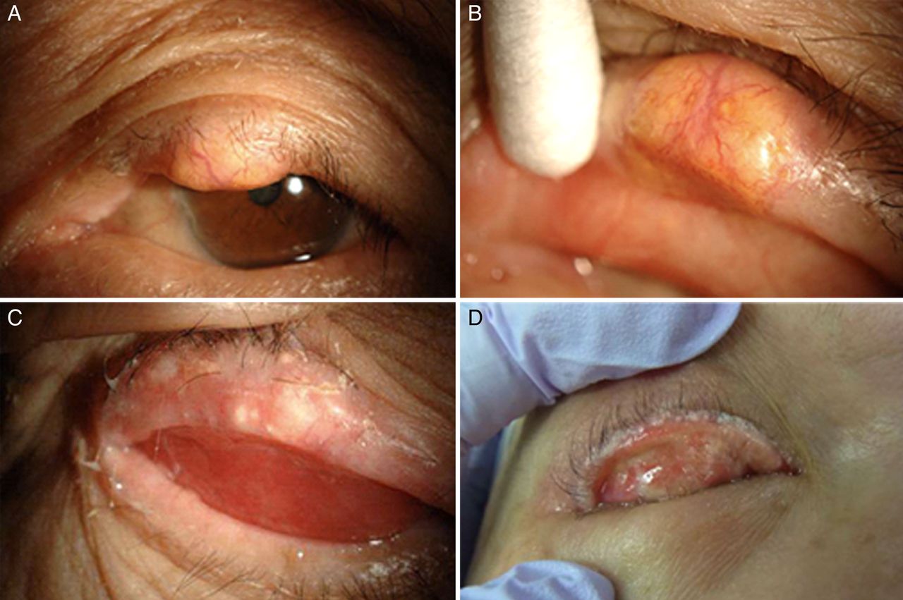
All but four cases (94%) presented initially as a distinct nodule or mass (figure 1). One mass was papillomatous, while the remaining were sessile in appearance. The remaining cases presented as diffuse thickening of the lid in three patients, and placoid appearance in one patient. All but two cases received the same clinical grading by the initial treating consultant and blinded grader. Both cases were initially noted as having a distinct nodule/mass but were blind graded as placoid and diffuse thickening. Nine cases were unable to be assessed by our blinded grader due to poor image quality or absent clinical pictures, but all of these were noted to have presented with a distinct nodule or mass. Sebaceous carcinoma was the correct initial diagnosis in 36 cases (57%). The remaining cases were clinically misdiagnosed as a chalazion (29%), blepharoconjunctivitis (5%), squamous cell carcinoma (3%), basal cell carcinoma (2%) or other (3%). The correct histopathological diagnosis was made in all but one case, which was misdiagnosed as a squamous cell carcinoma. This patient underwent surgical excision with reconstruction, and the final pathological report revealed sebaceous carcinoma with was totally excised.
(A–D) Clinical presentations of sebaceous carcinoma as a solid mass (A and B) and as diffuse pseudoinflammatory changes (C and D). The eyelid was everted for better visualisation.
Table 2 summarises the TNM stages for our patients. No patients presented with a T stage of T1 as all cases invaded the tarsus or lid margin. The majority of patients (60%) presented at stage T2aN0M0. There were eight patients (13%) with T3a or higher tumours and two patients presenting with regional nodal or distant metastatic disease.
TNM staging of patients with sebaceous carcinoma
The most common histological pattern was lobular, occurring in 25 cases (41%), followed by lobular and comedo (22 cases, 36%), comedo (10 cases, 16%), lobular and papillary (5 cases, 8%) and papillary (1 case, 2%). Pagetoid spread was demonstrated in 14 cases (21%), of which seven spread to the conjunctival epithelium, three to the eyelid skin and four spread to both. Twelve patients demonstrated only one quadrant of pagetoid spread, while the remaining two patients demonstrated two and four quadrants of spread.
The median size of the sebaceous carcinomas was 8 mm (range 2–37 mm). Margin controlled excision with frozen section was the most common primary treatment, performed in 51 (81%) patients. Wide margin excision (5 mm) without frozen section was performed in 10 cases (16%). One patient presenting with the largest sebaceous carcinoma measuring 37 mm (T3bN0M0) with orbital invasion required an exenteration. Two patients with stage T3aM0N0 tumours underwent map biopsies due to a clinical suspicion of intraepithelial spread. One of these patients presented with a diffuse lesion with four quadrants of pagetoid spread and received adjuvant topical mitomycin C. The second patient had a 20 mm diffuse lesion with two quadrants of pagetoid spread. Three patients received radiotherapy when surgical treatment was contraindicated. The first developed a local recurrence with extension into the orbital and maxillary sinuses and received radiation to the affected area. The remaining two patients underwent radiotherapy to metastatic disease (regional nodal in one patient, and regional nodal and distant in another) after being deemed unfit for surgery. Three additional patients with regional nodal metastases underwent lymphadenectomies.
Patients were followed up for a median of 4.2 years (mean 4.3 years, range 0.3–11.3 years). Complications secondary to surgery were noted in nine cases (14%) which included entropion (three cases), ectropion (two cases), ptosis (two cases), flap necrosis (one case) and graft necrosis (one case). Local recurrence occurred in four patients (table 3) who presented with initial tumour size varying from 4–20 mm. One patient developed multiple local recurrences at 15 months and 22 months following initial treatment, and required radiotherapy for the second recurrence, which involved the orbital and maxillary sinuses.
Cases of sebaceous carcinoma associated with local recurrence
Table 4 describes metastatic disease with respect to TNM stages. Five patients were found to have regional nodal metastases. Of these, two presented with regional metastatic disease, while three patients developed nodal metastasis during follow-up. All but one of our patients with regional nodal metastasis had tumours within the T2 category. There were two patients who presented with recurrent sebaceous carcinomas following initial treatment at different institutions. Both these patients subsequently developed regional lymph node metastases during follow-up after treatment of their recurrences.
Metastatic disease and correlation with TNM stage in patients with sebaceous carcinoma
Kaplan-Meier methods were employed to assess the relationship between T category and tumour outcomes; T3a tumours and greater were significantly associated with local recurrence (p=0.01) but not regional nodal metastasis (p=0.63).
Our only disease specific mortality occurred in a patient presenting with distant metastases (T3bN1M1), who died less than 1 month after starting heavy ion radiotherapy. One patient is alive with disease and all remaining patients are alive without evidence of disease at last follow-up.
Discussion
Our study represents the largest clinical review of Japanese patients with sebaceous carcinoma, and is also the largest series to date using the AJCC TNM staging system. We found that sebaceous carcinoma most commonly presents as a distinct eyelid mass in our Asian patient population. Additionally, in contrast to previous reports, tumours <10 mm in size were associated with regional nodal metastases.
Sebaceous carcinomas are classically associated with misdiagnosis and delayed treatment as they frequently masquerade as more benign conditions. Of our 63 cases, 57% were correctly diagnosed initially and all but one received the correct histopathological diagnosis after biopsy. These rates are significantly higher than previous reports. In a study of 60 patients, Shields et al9 found that sebaceous carcinoma was suspected in only a third of their patients and the correct histopathological diagnosis was made in only 50%. Even lower rates have been reported by Zucher et al,10 with only 18.6% of their patients receiving the correct initial diagnosis. Additionally, Leibovitch et al found that 75% of their cases were misdiagnosed histopathologically following biopsy.11 Our higher rates of correct initial diagnosis may reflect the increasing awareness of sebaceous carcinomas among Asian ophthalmologists and pathologists, given the possibly higher incidence of this condition in Asian countries.3 ,4 ,12
Previous series with predominantly Caucasian patients have demonstrated high rates of sebaceous carcinoma presenting with a diffuse pattern of eyelid thickening and the most common misdiagnosis reported within these studies was blepharoconjunctivitis.9 ,13 In contrast, 94% of our patients presented with a solitary mass or nodule, and 29% of our cases were misdiagnosed as a chalazion, compared with just 5% as blepharoconjunctivitis. This difference in presentation between Caucasian and Asian patients is also reflected in the literature, with chalazion being the most commonly reported misdiagnosis of sebaceous carcinoma from Japanese and Korean studies.12 ,14 This possible difference in clinical presentation may also be a factor in the lower rate of clinical misdiagnosis in Asian populations as compared with Caucasian ones.
The rate of intraepithelial spread within previous studies involving predominantly Caucasian patients has been reported as high as 51%, and these studies have suggested that map biopsies of the palpebral and bulbar conjunctiva should be performed routinely for sebaceous carcinomas.2 ,9 ,15 However, we found much lower rates, with just 21% of our cases demonstrating pagetoid spread histologically. Yoon et al14 reported even fewer cases of pagetoid spread within their Korean series, which occurred in only 8.3% of patients. Chao et al also demonstrated differing clinical features of sebaceous carcinoma depending on the presence of intraepithelial spread, with 72% of patients with pagetoid spread presenting with diffuse eyelid thickening and 43% of patients without pagetoid spread presenting with a distinct mass.16 However only 2 of our 14 patients with pagetoid spread presented initially with diffuse eyelid thickening. We used map biopsies only if there was clinical suspicion of epithelial involvement. Only two patients, both with stage T3aM0N0 tumours, underwent map biopsies. One of these patients developed multiple local recurrences at 15 months and 22 months after initial treatment. Our findings suggest that sebaceous carcinoma may exhibit different growth patterns between racial groups and therefore outcomes in our study may not be applicable to other ethnic populations.
The only previous study correlating TNM staging with outcomes for sebaceous carcinoma of the eyelid found that disease-specific survival was poorer among patients with T stage of T3a or worse.7 We had eight patients with T3a or worse tumours which included our only disease-specific mortality. Additionally, Esmali et al7 found that no tumours 9 mm or less were associated with lymph node metastases and that T category at presentation was significantly associated with nodal metastasis. However we had two patients with primary T2a tumours measuring 5 mm and 7 mm who developed nodal metastasis during follow-up. As a result, Kaplan-Meier analyses demonstrated that T category was not significantly associated with regional nodal metastasis. Therefore while Esmali et al recommended sentinel node biopsy or strict node surveillance for tumours T2b or worse, our findings suggest that all patients with sebaceous carcinoma require careful nodal surveillance and follow-up, as even tumours of T stage of T2a are associated with regional nodal metastasis. Additionally, while Esmaeli et al found that T-category was not associated with local recurrence, we found that tumours of T3a and above were significantly associated with local recurrence. However our findings are limited by our small numbers and further studies investigating the prognostic value of the TNM staging system are required to better evaluate the role of the TNM classification in eyelid sebaceous carcinoma.
The majority of our patients underwent margin controlled excision with frozen section, and despite some debate regarding the advantages of Moh's microsurgery compared with frozen section, both are widely accepted options for definitive surgical treatment.1 Frozen section monitoring has previously been associated with false negative rates in 25% of cases, and for this reason, many centres advocate the use of fast-tracked permanent paraffin-embedded sections and delayed closure.13 ,17 However, in centres where pathologists are experienced with the use of frozen section for margin control, the rate of false negatives is low, and recurrence rates appear to be comparable with permanent section.1 ,14 Frozen section was the preferred form of margin control at our institution, which may be reasonably effective in the short-term, as we had a recurrence rate of 8% (4/51) during a median follow-up of 4.2 years. However, the differences in growth patterns for patients of other ethnicities should be taken into account when considering the method of margin control most suitable.
Our study has a number of limitations which warrant recognition. The retrospective nature of this study precluded the collection of additional clinical information which may have provided incremental insights. Previous reports have documented local recurrence rates occurring at a median of 20 months, and although our median follow-up period was 4.2 years, further recurrences may occur beyond this follow-up period. Our small numbers of local recurrence and regional nodal metastasis should also be taken into account when considering the results of our statistical analysis. Additional follow-up is required to assess the recurrence rate within our patient cohort and assess the efficacy of frozen section control in this context.
Conclusion
The clinical presentation of sebaceous gland carcinoma in our Japanese population consisted largely of eyelid masses with a blepharoconjunctivitis-type presentation being rare. This appears to differ from many Caucasian reports and is suggestive of a difference in clinical presentation between racial groups. Additionally, we found lower rates of histological intraepithelial spread compared with previous Caucasian series. In contrast with previous reports, we found that T2a tumours smaller than 10 mm were associated with the development of regional nodal disease and T3a tumours and greater were significantly associated with local recurrence but not regional nodal metastasis. The local recurrence rate for patients treated with frozen section controlled excision was 8% (4/51) at a median follow-up of 4.2 years. Further prospective controlled studies with adequate follow-up will be required to determine the efficacy of standard frozen section controlled excision as compared with fast-tracked paraffin control or Moh's surgery.
References
Footnotes
-
Contributors All authors fulfil the criteria for authorship and there were no additional contributors. AW (first author) was responsible for the identification and management of cases in our series.
-
Competing interests None.
-
Ethics approval Kyoto Prefectural University of Medicine Hospital Ethics Committee and Seirei Hamamatsu Hospital Ethics Committee.
-
Provenance and peer review Not commissioned; externally peer reviewed.
Linked Articles
- Correction