Article Text

Statistics from Altmetric.com
Background
Patients with low vision typically have reduced visual acuity and a significant loss of contrast sensitivity, often in combination with visual field loss. These impairments cause a number of disabilities including difficulty with reading, writing, recognising faces, watching television, orientation and mobility, and completing activities of daily living. In a recent large scale survey of providers of low vision services, Elliott et al 1found that for elderly patients with low vision, the primary objectives identified at low vision assessment were to obtain help with reading and with vision oriented performance of daily living activities. Secondary objectives commonly include obtaining help with watching television, mobility and independent travel, and hobbies. In many cases, these objectives can be met by the prescription and use of conventional low vision aids (LVAs)—that is, optical devices providing magnification in order to compensate for reduced visual acuity, while contrast is maximised with local task lighting. LVAs are, unfortunately, highly task specific, and the patient may need several different aids to deal with a variety of identified requirements. Recently, however, alternative devices have been introduced which offer a number of distinct advantages over conventional LVAs in low vision rehabilitation. The purpose of this review is to describe these devices and to discuss both their current and future potential in comparison with existing technology.
Existing technology for low vision rehabilitation
Contrary to early work suggesting that LVAs are often limited in their effectiveness,2 3 more recent studies indicate that low vision rehabilitation and the prescription of LVAs can offer considerable benefit to the visually impaired, with at least one device being used by between 80% and 91% of patients provided with an LVA.4-8 It is recognised, however, that some patients do not continue to use LVAs following their dispensing. While some users discontinue with an LVA because of a change in vision, research has shown that a patient’s age and the visual acuity achieved with devices are not predictive of their continued use.4 6 8 Some users discontinue using LVAs because of the ergonomics of the device, frustration with the optical limitations, or in some cases users obtain another device or solution to the problem.6 A recent survey of LVA use by US veterans6 revealed that these users would like to see a range of improvements in LVAs including a wider field of view, ability to see “greater detail”, automatic focus, a desire to see things nearer/further away, and additional image brightness. A much smaller percentage of this group of veterans also wanted the devices to be “less noticeable”. There are, therefore, limitations associated with conventional LVAs.
When conventional LVAs produce insufficient magnification or are too restricted to provide for a sustained near task, closed circuit television (CCTV) technology or other electronic devices may be helpful. Electronic systems have the advantage of being able to provide high magnification with a relatively large, aberration free field of view. The eye to device working distance is typically larger than for an optical magnifier, allowing binocular viewing from a natural, comfortable posture. Comparing patient performance with conventional LVAs and CCTV systems is difficult, since not all patients are equally familiar with both. Goodrich et al 9 tested 96 US veterans who had been CCTV users for at least 2 years. They found that 50% of their patients used both conventional LVAs and a CCTV, often in combination, with an optical device being used to find an item of interest and the CCTV being used for subsequent detailed viewing. Other applications of conventional LVAs were for short term “spotting” tasks or in situations where portability was important. For those patients who had an up to date optical LVA, the reading speed was tested with both this aid and a CCTV and found not to be significantly different, although the duration of use was about three times longer with a CCTV.
Once a device such as a CCTV reaches the commercial marketplace, there appear to be two ways in which it can develop— either “up” in the technology scale, incorporating ever more sophisticated features, or “down” in an attempt to make the device more accessible and affordable. Despite significant developments in CCTVs,10including the development of head mounted devices (HMDs) such as the Magni-Cam (Innoventions Inc), these devices are usually restricted to helping with a limited range of near tasks, with many of the innovations in CCTV systems typically going “down” the technology scale. For example, the use of hand held cameras,11connection to domestic television (Scantec), and adaptations to a “camcorder”12 are developments which have usually been driven by economic considerations. While limiting the options and versatility, these developments sometimes offer practical advantages and have been presumed to be adequate technically to achieve the same purpose as a conventional system. Despite the attempts to bring CCTV systems to a larger audience, however, it appears that only a small minority of people with a visual impairment use this technology.
An emerging technology
Until recently, there has been a greater technological effort in developing aids for the blind rather than for those with low vision.13 However, recent advances in optoelectronics and video technology have permitted the development of new low vision rehabilitation devices (for example, the low vision imaging system (LVIS), formerly known as the low vision enhancement system), which in contrast with other devices, have been designed to be used for a variety of tasks. The LVIS, developed at Johns Hopkins University,14 15 is a mains/battery powered video HMD, equipped with autofocus camera, variable magnification optics, and contrast enhancement electronics (that is, image processing capability). The system contains two video cathode ray tubes mounted in the device’s temple arms on each side of the head. Video screen images are magnified by aspheric lenses and imaged to the front of the eyes by mirrors and beam splitters (Fig 1). The device is equipped with two monochrome charge coupled device cameras mounted in front of the eyes, approximately on axis with the line of sight (and providing an unmagnified binocular field of view for orientation) and a third, centre mounted zoom camera (providing the same image to both eyes with variable magnification). This latter variable focus camera can tilt down by up to 45° for near vision/downgaze. In standard configuration, 10.5× magnification is available at near, although an auxiliary flip up reading addition lens is necessary to achieve higher near vision magnification.

Schematic diagram of the LVIS. A magnified intermediate image of each cathode ray tube (CRT) screen is formed by an aspheric triplet lens system. Light from the temple arm CRT is directed by the folding mirrors to the 50/50 beam splitter in front of the eye. The curved mirror images the exit pupil of the optical system in the plane of the subject’s pupil and displays a 50° × 40° image of the CRT screen. A refractive correction, if necessary, can be inserted between the CRT and the aspheric triplet (redrawn after Massof et al 15).
The head mounted design frees the user’s hands. The magnification, contrast, and brightness are under user control (Fig 2), and, unlike conventional LVAs, can be varied for a wide variety of tasks, in order to provide the optimal conditions for each user/task. For example, image contrast can be reversed. The LVIS also features the facility to view a television via direct video feed.

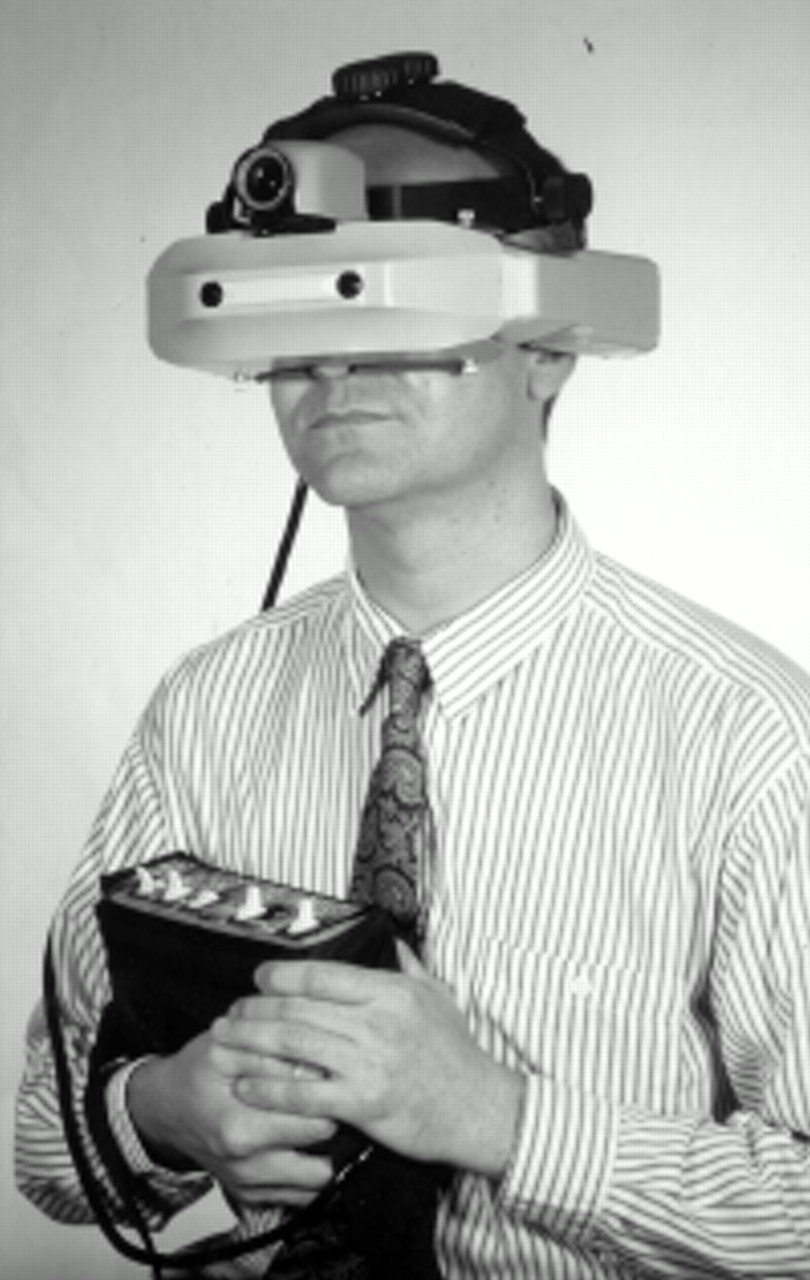
Photograph of the LVIS. The two monochrome charge coupled device cameras mounted in front of the eyes provide an unmagnified binocular field of view for orientation and the third centre mounted zoom camera provides variable magnification. The control unit can be worn as a “belt pack” and consists of a series of switches (for example, to enhance contrast, change contrast polarity, or “toggle” between orientation and zoom cameras). The function of the switches must be learnt, since these cannot be seen when the unit is worn.
A monocular video magnification HMD, known as the Aurora imaging system, has also been developed by the manufacturers of the LVIS but at the time of writing is currently not available in either the USA or the UK. This system has a smaller, lighter headset than the LVIS, but retains the variable magnification and contrast enhancement capabilities. Another system, known as the V-max (Enhanced Vision Systems), is also marketed in the USA and has recently been introduced in the UK. This system is also a battery powered head mounted unit, but differs in that it uses a colour camera and liquid crystal displays. Image processing is available in the form of edge enhancement technology and contrast reversal. The control box is smaller and simpler than that of the LVIS. The V-max also features a docking stand, permitting the device to be used like a conventional CCTV when connected to a monitor. Table 1 summarises the principal features of the two video magnification HMDs currently available in the UK.
Specifications and features of the two video magnification head mounted devices currently available in the UK (manufacturer/supplier data)
This video magnification technology has the potential to offer considerable improvements to a patient’s visual functioning and the ability to perform activities of daily living when compared with the use of conventional LVAs and would appear to offer solutions to some of the issues raised by users of conventional devices.6 In summary, the advantages include: (1) head mounted design means that the device leaves the hands free; (2) flexibility of variable magnification over a wide range of viewing distances means that the device might be used for a range of tasks; (3) contrast enhancement and reversal permit individuals to select optimal images for different tasks; (4) automatic video gain control permits the maintenance of a constant screen luminance, a feature of particular use for subjects with adaptation/glare difficulties (for example, retinitis pigmentosa); and (5) the possibility for a wide field of view with binocular vision.
Disadvantages include the weight of the headset, the appearance, the cost, the time consuming assessment and fitting procedures (arguably less problematic with the V-Max system than with the LVIS), the complexity of operation and potential problems with motion sickness and claustrophobia. In addition to these disadvantages, our initial impressions of an early (but commercially available) LVIS indicate slow and inaccurate focus of the autofocus camera and limited resolution of the video display. The systems are unsuitable for patients with serious head/hand tremors or those patients physically unable to operate the controls or support the headset.
Evaluating the new technology
In describing the development of the LVIS, Massof and Rickman14 describe a phased programme, in which several generations of the device are made available in quick succession. This process seems appropriate, since a design feature or patient selection criterion which appears reasonable when the device is still a theoretical concept may prove non-optimal when tried in practice. Therefore, a comparative trial of the new technology at a very early stage, when one might become committed to an ineffective strategy, is arguably not ideal. However, once the technology and the basic methods of assessment are more established, then it becomes important to make comparisons with existing modalities. The danger in failing to evaluate the systems at this stage is that changes in subsequent versions of the device are more likely to be driven by technological developments without due consideration as to whether the features provide improved performance.
Massof and Rickman14 describe their primary goal in developing the LVIS as solving “the functional problems encountered by patients with low vision”. A preliminary case series of 43 users reported by Maino et al 16indicates that the systems are used for a range of different tasks including reading, watching television, computing, sightseeing, typing/writing, and doing repairs (for example, in the home, to the car, etc). Thus, it would appear that this device can meet many of the patient objectives identified during low vision assessments.1 Massof and Rickman14 also suggest that the new technology should “compete effectively with alternative technology”, to which we would add “and existing devices”. Preliminary evaluations of the LVIS indicate both improved visual function (acuity and contrast sensitivity) and improved task performance when compared with the patient’s habitual spectacle correction or optimal conventional LVA.16-18 The authors are not aware of published reports in academic journals describing the effectiveness of the other video magnification HMDs. A study of 60 patients with low vision by Rohrschneider et al 17 found an “average” improvement of “eight log steps” with the LVIS (that is, eight lines on a logMAR visual acuity chart) compared with the patients’ habitual spectacle correction and up to “three log steps” compared with conventional telescopic devices. Arguably of more importance, however, is the improvement in contrast sensitivity with the LVIS, an improvement not available with conventional LVAs. Rohrschneider et al 17 suggest improvements from “0 to 16 steps” using the Pelli-Robson low contrast letter chart but do not clarify in their report what is meant by an improvement of “16 steps”. While this finding appears to suggest that it is possible for some patients to obtain an increment in contrast sensitivity which spans the entire dynamic range of the chart (that is, 2.40 log units), a study of 46 subjects with low vision suggests that the average improvement in contrast sensitivity is ∼0.50 log units (Massof, personal communication).
Further research is needed in order to determine whether these improvements in visual functions (that is, visual acuity, contrast sensitivity, and glare sensitivity) translate into improvements in task performance and quality of life for the users of the systems, since it is recognised that visual function may be misleading as a measure of “success” with a low vision device.4 6-8 19 Despite these encouraging preliminary findings, the authors are not aware of any controlled trial to determine the benefits of these systems. Thus, it remains to be seen whether the benefits reported in selected case series have wider implications for the low vision population. More difficult to verify and define, yet just as important for long term success, is the requirement of acceptability to the patient, in terms of appearance, comfort, and ease of use. Case reports suggest that motivated subjects can use the system for 8–10 hours a day20 and yet, in the study by Rohrschneideret al,17 the majority of patients when questioned could not imagine using the system regularly. This apparent contradiction merits investigation to ascertain why a particular device may not be appropriate for some patients. It is important to know in advance whether an expensive electronic aid which requires a lot of practice would be suitable for the patient. A previous evaluation of the effects of training and practice with conventional LVAs and CCTVs concluded that subjects require about 2–3 weeks in order to reach peak reading speed.21 It seems likely that the more diverse range of tasks permissible with the more complex video magnification HMDs will necessitate an even longer period before optimal performance levels are reached.
Goodrich et al 9 were among the first to point out that the clinical prescribing of LVAs is best accomplished by comparing a patient’s task performance with different devices, since it is not possible to use simple quantifiable clinical variables to predict performance. In discussing successful use of CCTV systems, they note that the factors which determine performance are unknown. Goodrich et al 9suggested at the time that the “relatively intangible” area of motivation be explored and that muscular strain may be a limiting factor in reading duration with conventional LVAs; these factors have yet to be investigated. Only rarely considered in respect of the adaptation to using optical magnifiers (since they are typically used by a stationary patient) is the adequacy of retinal image stabilisation during head movements. For a normal subject, whenever a head movement occurs there should be an eye movement of equal speed but in the opposite direction. This movement is generated by the vestibulo-ocular reflex, which if functioning perfectly, would have a gain of 1.0 (that is, eye velocity/head velocity = 1.0). Measured in darkness, the vestibulo-ocular reflex provides a substantial measure of stabilisation of the retinal image during head movements. In the light, visual mechanisms including pursuit and optokinetic response interact synergistically to optimise the gain of compensatory eye movements. Even this visual vestibulo-ocular response is limited in its capacity to produce effective image stabilisation at higher rotational frequencies and velocities. When viewing through a magnifying system, retinal image motion is magnified, and the visual vestibulo-ocular reflex gain must be equal to the magnification to stabilise the retinal image. Dynamic visual acuity, the acuity during imposed head motion, is a measure of the effectiveness of retinal image stabilisation. In normal people, the visual vestibulo-ocular reflex functions adequately during head motion to limit retinal image instability, making dynamic visual acuity independent of head velocity. If the visual vestibulo-ocular reflex gain is insufficient, then there will be retinal image slip, and if this slip is greater than 2 deg/s then dynamic visual acuity falls. Although the telescope magnification is helping to mitigate this loss of vision, the lack of compensation of the eye movements for head movement is such that when head velocity exceeds 20 deg/s, all telescopic spectacles actually cause a decrease in dynamic visual acuity, regardless of their magnification.22 These head velocities are typical of those encountered during normal walking, and completely negate the intended visual advantage of telescopic magnification. In fact, Demeret al 23 found that unsuccessful telescope users made more spontaneous head movements, and had much poorer dynamic visual acuity than successful users, thus compromising their vision during normal wear of the devices. As well as decreasing acuity, this retinal image slip can create severe sensations of motion sickness in susceptible patients: sometimes to the extent that such telescopes cannot be used. Anecdotal reports indicate that some patients experience a similar problem with the video magnification HMDs.
This discussion would seem to suggest that magnifying systems for constant wear would never be effective, yet this is plainly far from the truth, since there are some patients who have successfully worn “contact lens telescopes” for extended periods. It has been suggested that the retinal image motion created by these Galilean telescopes (produced by a negative contact lens, usually >−30.00DS, and a positive spectacle lens, typically >+15.00DS), “would pose insurmountable obstacles and render the use of telescopic systems impossible”.24 In fact, nowhere in the literature on contact lens telescopes does it suggest that retinal image slip is a cause of failure in adapting to the systems. There are several possible explanations for this failure to adapt. Firstly, there is a great deal of individual variability in dynamic visual acuity, even among normal subjects who had the same static acuity,22 and it may be that successful wearers are those with better dynamic visual acuity (although whether this reflects a more competent visual vestibulo-ocular reflex or a viewing strategy which allows them to better interpret a moving retinal image is not known). Secondly, with even relatively short periods of exposure to a magnified retinal image, the visual vestibulo-ocular reflex appears to adapt,23although the optimal way in which to manage this adaptation is unknown. Many questions remain unanswered: how long does it take to adapt and is a single long exposure equivalent to several shorter periods? Does the adaptation disappear if not reinforced by regular exposure? Can subjects switch instantly between the visual vestibulo-ocular reflex gain required for magnified and that required for unmagnified viewing? Finally, it is interesting that many of the successful contact lens telescope wearers are patients with congenital nystagmus, and they are also more successful users of conventional telescopes than non-nystagmus subjects.25 This success could be because subjects with nystagmus have an adapted visual system to deal with large amounts of retinal image slip.
One further point to mention in relation to the use of a HMD is the considerable interest in both the scientific literature and the popular press concerning possible adverse effects of HMDs (usually “virtual reality” displays). Peli26 has shown that for normal subjects there are no clinically significant changes in functions such as accommodation or binocular vision following 30 minutes’ use of a HMD. Any measurable changes which do occur do not appear to be specific to the use of the HMD, since they would also be found after use of a desk top display, although using the former produces vague subjective complaints of eye strain and discomfort in some subjects.
To summarise, the available evidence would indicate that in their present format, the current video magnification HMD systems are likely to be suitable for only a minority of patients with low vision. Motivated, mentally alert subjects with a positive attitude towards rehabilitation (including those who are successful with conventional devices) seem to be the most likely to be successful, although it remains a priority for researchers to identify the patient factors which best predict successful use.
Future developments in image enhancement
The technology employed in the devices described has already been superseded. Further developments are likely to address the principal limitations. Future devices are likely to be smaller and lighter and display images of improved quality. However, it is interesting to speculate on what constitutes the optimum display characteristics for low vision. The argument has been advanced by Leat and Rumney27 that optimising image quality may lead to wasted resolution—that is, it is not worthwhile to design a system which faithfully transmits spatial frequencies in the image which cannot be resolved by the user. It might be argued that some sacrifice in “quality” (as perceived by normally sighted users) would be preferable if it resulted in a less expensive, larger, brighter, or lighter weight display, for example.
One of the major advantages of electronic systems over optical systems is that the image can be enhanced in order to improve the contrast (and hence visibility) of images. In current CCTVs, this enhancement is a homogeneous alteration for printed text (that is, non-customised), although varying and/or reversing the contrast can be an individual choice. Within the video magnification HMDs, variable contrast enhancement can be applied to a wide range of visual tasks (for example, face recognition) but it is still not customised for the individual. Customised contrast enhancement is, however, feasible with these electronic systems. To represent the situation very simply, the loss of contrast detection ability caused by ocular pathology can be represented as the effect of a linear filter. The ratio of the contrast sensitivity function of the impaired person to that of a normally sighted individual measures the filtering effect of the defect. Applying this filter to the perceived image would simulate the effects of the impairment, which typically involve high spatial frequency loss: the inverse of this filter applied to the abnormal contrast sensitivity function would return perception to normal. This process would be one way of creating image enhancement, although there are practical difficulties. Firstly, substantial high frequency noise in the image is emphasised by the enhancement of these frequencies. Secondly, if the contrast variations in the original image use most of the available dynamic range then the required “multiplication of the contrast” cannot be achieved, although if attempted (and the resulting image rescaled to fit the available range) there will actually be a loss of contrast at some frequencies. However, since there are some (high) spatial frequencies which will not be visible to the low vision observer (regardless of the enhancement) the image can be low pass filtered to remove these spatial frequencies.
These image enhancement strategies relate to theobserver, and could be customised to the requirements of an individual patient, changing as the pathology progressed. It is equally possible, however, to consider the spatial frequency characteristics of the imageitself (this time measured in cycles per object), and choose to amplify only those spatial frequencies known to be important for recognition. In the case of faces, for example, it is known that the most useful region is around 20 cycles per face.28 The optimum bandwidth will of course be different depending on the particular recognition or identification task to be attempted, suggesting that the enhancement would need to be adjusted accordingly. Perhaps the situation need not be as complicated as this would suggest: one would expect that a detection task would depend on low frequencies, whereas recognition demands high frequency detail, so selecting between the two alternatives of either low or high spatial frequency enhancement may be sufficient.
Peli and co-workers have found improved performance of visually impaired patients for recognition of faces, expressions, and other details on still photographs29 and video films.30 Images enhanced in such a way will present a grossly distorted appearance to the observer, however, and patients may prefer not to see objects in this way, despite the improvement in performance.
As noted above, contrast enhancement has only been utilised in CCTVs in a limited way (that is, non-customised uniform enhancement), despite the fact that possible approaches were considered some 20 years ago.31 Typically, the image on the CCTV screen has a higher contrast than the printed page from which it comes, and its contrast can be reversed which seems to be preferred by many users. It is surprising that the technique described above has not been developed further in view of the importance of contrast to reading. For example, the work of Whittaker and Lovie-Kitchin32 33 suggests that image contrast needs to exceed the patient’s threshold by a factor of at least 10 if reading is to be fast enough to “read for pleasure”. Peli et al 30 did not find any improvement in reading performance for text which had been custom enhanced in this way, contrary to the results of Lawton,34 35 who reported dramatic increases in reading speed. Perhaps these somewhat equivocal findings may explain, at least in part, why customised contrast enhancement has not been advanced in CCTV systems.
Traditionally, CCTVs have used black and white displays, with a colour system typically used only for special applications, such as viewing maps and in education. Recent years have seen the advent of systems which allow the user to display original monochrome images (of text, for example) in contrasting colours, but there is little evidence of the usefulness of this approach. For reading text it appears that maximising luminance contrast is the only way to optimise performance, although patients may subjectively prefer to introduce colour contrast as well. Unfortunately, the quantitative objective evidence of performance improvement to support these preferences does not exist currently.36 Presumably a patient’s preference for colour contrast is related to psychological or aesthetic benefits, or improved “visual comfort”, providing that high luminance contrast is still available to maintain reading speed.
It is interesting to note that of the two video magnification HMDs currently available in the UK, one is monochrome and the other colour. While there is no strong evidence in support of the use of colour displays for reading, it is possible that colour improves performance in tasks which require object recognition, since patients with low vision will not necessarily have access to subtle shape and texture information which might render colour redundant. Wurmet al 37 carefully controlled for luminance contrast in their images, and found that colour did improve the speed and accuracy of naming familiar objects (food items). This finding led them to suggest that colour contrast is a useful practical strategy to aid the performance of a recognition task, but that the practical benefit of colour versus monochrome displays needs to be investigated experimentally.
Den Brinker and Beek38 remind us that the fundamental design of the human visual system is the combination of a small retinal region of high acuity with an eye head body motor control system to achieve sharp images of our whole environment. This system requires the precise direction of the fovea to objects of interest, once these have been detected and located. This switching of attention from detection and location to the processing of the required information, is extremely difficult with any LVA, notwithstanding the effects of the visual loss itself. The way in which this task is accomplished, and the optimum design of an LVA to facilitate the process, has never been fully explored. The bioptic design of some spectacle mounted telescopes makes use of this concept, as do the new video magnification HMDs and the zooming facility (variable magnification) on most CCTV cameras. Whether the two images are best presented successively or simultaneously has not been investigated. It is not known whether this facility is desirable for any or all of the tasks which might be attempted with a magnifier, although den Brinker39 has shown that a pointer indicating the camera location on a CCTV is beneficial for speeding search and reducing nausea.
The other image manipulation which might be attempted is spatial remapping. Unlike contrast enhancement, this form of enhancement can only be done on a customised basis, and would require real time monitoring of eye position, thus adding to the complexity and bulk of the system. Parts of the image which fall into a scotomatous area can be repositioned onto adjacent functional retina. The image obviously becomes somewhat distorted by this process, since the extra information has to be squeezed into space already occupied, and so partial remapping may be an acceptable compromise. In simulations with such a system, subjects have still read much more slowly than normal40: any improvements which were produced were very small, and the possible real life usefulness of such changes needs to be evaluated.
Conclusions
While this emerging technology has limitations at present, it is possible that such devices could make a significant impact on the rehabilitation of low vision patients in the future. Despite likely advances in this technology, it is timely to evaluate the potential of the systems in order that their diffusion can be rationalised. With the high media coverage and the allure surrounding new technologies ever present, understandably there has been much interest in this technology among the visually impaired. There is, therefore, a pressing need for a prospective controlled trial of these devices versus conventional LVAs in order to determine the extent to which the new technology can contribute to improved quality of life for the low vision population. It is also necessary to establish appropriate methods for the evaluation of the emerging low vision technologies. We propose that any such evaluation should not rely solely on the traditional clinical measures of visual function but should include a range of valid quality of life measures. Within the present evidence based climate, there has been an inertia in directing funding towards low vision rehabilitation and consequently it seems likely that for the immediate future at least, both the cost and the time consuming nature of assessment and training prevents rehabilitation with these new systems from being encompassed within existing NHS low vision services.
Acknowledgments
Note: Both the LVIS and the V-Max are marketed in the UK by Visionary Imaging Systems Ltd. The authors have no financial interest in either this company or the US manufacturers of the systems discussed.